
Weak Contrast in Live-Cell Imaging: Solving It with Inverted and Phase Contrast Microscopes
A culture flask has been on the stage three times this week. The cells are alive, the medium is fresh, the focus is as sharp as the stage allows — and the image is still a pale grey haze with no readable cell boundaries.
That situation is the most common contrast complaint in biotech and medical laboratories, and it is rarely a hardware failure. Weak contrast in live-cell imaging usually comes from a mismatch between three things: the optical geometry of the specimen, the alignment of the phase contrast light path, and the observation mode the objective was actually designed for. Correct those three, and the same instrument that produced grey haze produces defined cell edges, internal structure and usable morphology.
Chongqing Scope Instrument Co., Ltd. is a laboratory and medical instrument manufacturer and supplier located in Chongqing, China, offering microscopes, metallographic equipment, hardness testers and medical manikins, with OEM and ODM services. Three of its platforms cover most of the contrast problems described here: the inverted BDS500 and BDS400 microscopes for vessel-based live-cell and tissue work, and the upright BK5000-PH phase contrast microscope for fixed specimens. The sequence below follows the diagnostic order that actually resolves the problem — specimen and light path first, hardware second.
What Weak Contrast Actually Looks Like
Contrast failure in live-cell and tissue imaging is a family of symptoms, not a single fault. Naming the symptom narrows the cause.
- Cells remain invisible until the culture is nearly confluent, then appear as a flat grey sheet.
- Cell outlines are visible at 4X or 10X but disappear when you switch to 20X or 40X.
- Structures are visible but surrounded by bright halos or a glowing rim.
- One part of the field is bright and another is dim, even after careful focusing.
- Tissue sections show their bulk outline but none of their internal detail.
- The image looks acceptable through the eyepieces but flattens out in the camera capture.
The reason behind most of these symptoms is physical rather than mechanical. Bright field contrast depends on the specimen changing the amplitude of transmitted light — absorbing or scattering it — so that intensity differences appear in the image. Living cells are mostly water and protein: they change the phase of transmitted light far more than its amplitude. A thin, unstained, transparent specimen therefore produces almost no intensity difference in a bright field image.
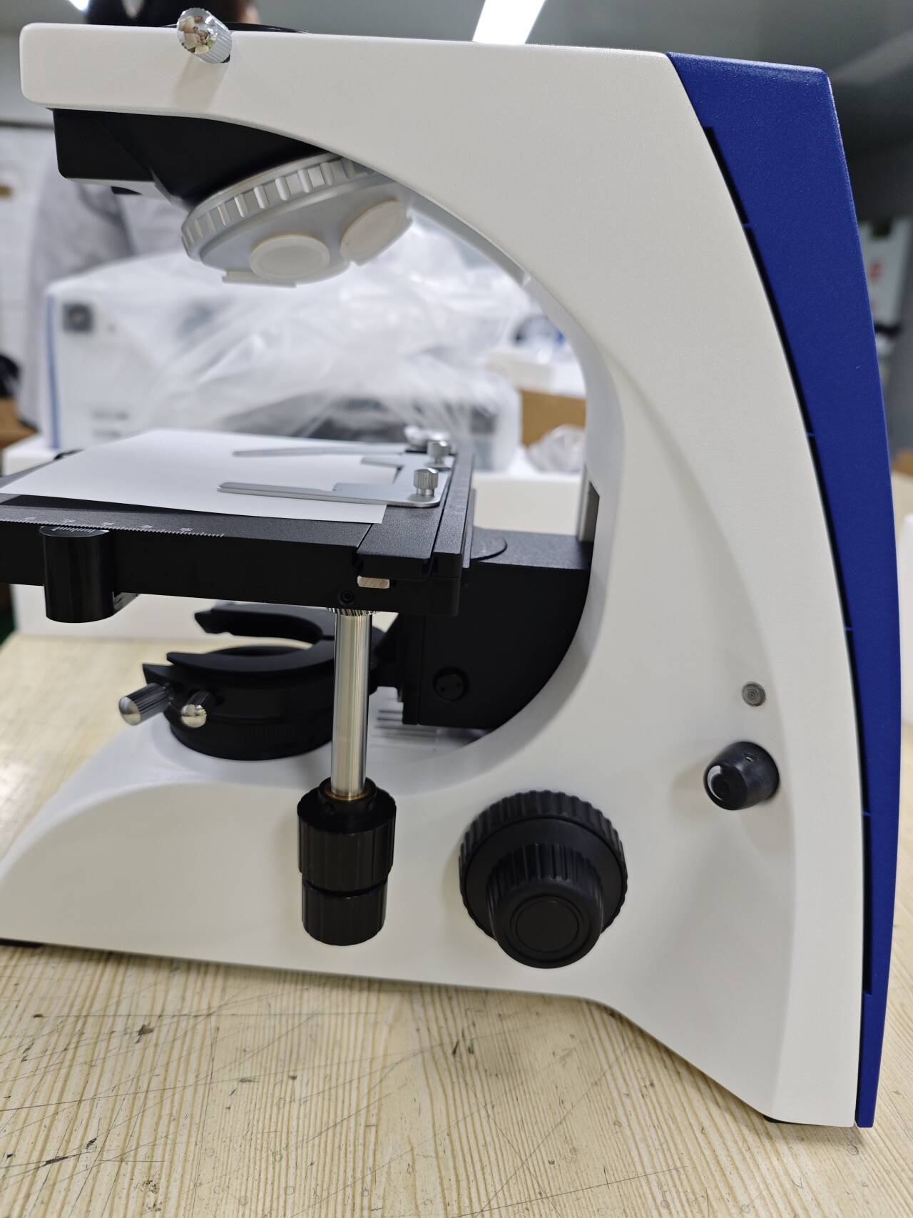
Phase contrast optics convert that phase difference into a visible intensity difference. An annular diaphragm in the condenser projects a ring of light through the specimen, and a phase ring inside the objective shifts the diffracted light so that regions of different optical path length appear at different brightness levels. Annulus and phase ring have to match in position and in magnification. When they do not, the conversion fails, and the observer sees a bright, even, low-information field — often with bright halos added on top.
Why Live-Cell and Tissue Work Still Depends on Optical Phase Contrast
Transmitted-light optical microscopy remains the default observation method for routine cell and tissue work, and the market data reflects that. Market research published in 2026 valued the global microscope market at approximately USD 11.9 billion in 2025, with optical microscopes accounting for approximately 37% of that market (Market Research Future, 2026). Trade data points in the same direction: global trade in microscopes under HS code 9011 reached USD 3.36 billion in 2024, with China the third-largest exporter at USD 430 million in export value (Observatory of Economic Complexity, 2026, drawing on UN Comtrade and national customs data).
Three practical consequences follow for laboratories and for the distributors who supply them.
- Most daily observation targets are transparent and unstained. They cannot be made readable by increasing magnification, because magnification enlarges an image that carries no contrast information.
- Contrast is a configuration decision. The same microscope body can be assembled around different observation modes; the mode and its matching optics, not the magnification, determine whether an unstained sample is readable.
- Because these platforms are widely traded, buyers can compare configuration detail before purchase — condenser type, annular spot coverage, working distance, and the documentation that travels with the instrument.
Step-by-Step Diagnostic Breakdown
Work through the three checks below in order. Each one eliminates a class of cause, and most laboratories find the answer within the first two.
Diagnostic 1 — Sample Thickness and Working Distance
If the specimen sits in a culture flask, a dish or a multi-well plate, light must pass through the vessel base before it reaches the objective. That geometry affects both the objective and the condenser. The symptoms of a working-distance mismatch are consistent: the vessel base comes into focus but the cells do not, or illumination is uneven across the field, or fine structure appears only at the very centre of the image.
Two checks resolve it.
- Confirm the configuration is inverted for vessel-based work. In an inverted microscope the objective sits below the stage and the condenser above — the geometry vessels are built around. The inverted BDS500 and BDS400 both provide a 54 mm culture dish holder and accept long working distance objectives: BDS500 carries LWD Infinity Plan Objectives at 4X, 10X, 20X and 40X for phase contrast and bright field, and BDS400 carries LWD Infinity Phase Contrast Objectives at 10X, 20X and 40X.
- Confirm the condenser is matched to that geometry rather than to a thin slide. BDS400 uses a long working distance condenser with N.A. 0.3, designed to deliver an even light cone through the vessel base. Where the limiting factor is the specimen itself rather than the vessel, reduce the thickness of the section or choose the objective that provides the required depth of field instead of pushing magnification higher.
Diagnostic 2 — Phase Contrast Condenser and Annulus Alignment
Phase contrast fails silently when the annular diaphragm in the condenser does not correspond to the phase ring of the objective in use. The field stays bright and even, but nothing separates from its background.
- Match the annulus to the objective magnification. BDS500 uses a 4-hole rotating disc phase contrast condenser with annular spot positions for 4X, 10X, 20X and 40X, so the correct annulus is selected by rotating the disc. BDS400 provides annular spots for 10X, 20X and 40X with its long working distance condenser. The upright BK5000-PH pairs infinity phase contrast objectives (Plan 4X, 10X, 40X, 100X) with a quintuple-hole turret Abbe condenser.
- Centre the annulus on the phase ring. Decentration typically appears as uneven contrast across the field or a bright rim on one side. This is an alignment step, not a repair.
- Set illumination before judging contrast. Köhler illumination is the baseline: centre the lamp, adjust the field diaphragm, then evaluate. BDS500 uses Köhler illumination with a 12V/50W halogen source. If the lamp is not centred, no annulus position will look correct.
Diagnostic 3 — Objective-to-Observation-Mode Matching
An objective is swapped but the observation mode is not. This is the most expensive silent error in routine microscopy, because the instrument is blamed for a configuration decision.
- Phase contrast objectives contain a phase ring; bright field objectives do not. A bright field objective with the annulus engaged produces a dim, flat image. A phase objective with the annulus disengaged produces a bright but low-relief image. Both look like weak contrast, and neither is a fault in the microscope.
- Check where the light is being sent. BDS500 and BDS400 provide light distribution of 100:0 and 80:20 between the eyepieces and the camera port. When the split favours the camera, the visual image appears dimmer than expected; when the full photographic light budget is needed, an adapter for digital photography routes 100% of the light to the photography port.
- Use the supplied filters deliberately. Blue and green filters ship with both inverted platforms and are intended for balancing transmitted light. Leaving them out, or combining them arbitrarily, shifts perceived contrast.
- Record the pairing. A simple log of objective magnification and annulus position prevents the mismatch from recurring when several people share one instrument.
Detailed Solution: Matching the Platform to the Specimen
Once the diagnosis confirms a genuine configuration gap rather than a setup error, the platform choice follows from three facts about the specimen: whether it sits in a vessel or on a slide, how thick it is, and whether it is living or fixed.
BDS500 — Inverted Platform with a Rotating Disc Phase Contrast Condenser
BDS500 is an inverted laboratory microscope designed for cell culture, biology and laboratory research. Its most relevant feature for contrast work is the 4-hole rotating disc phase contrast condenser, which places annular spot positions for 4X, 10X, 20X and 40X in a single rotating disc, so switching phase contrast magnification becomes a rotation rather than a change of condenser insert.
- Head: trinocular
- Eyepiece: WF10X/22mm
- Objectives: LWD Infinity Plan 4X, 10X, 20X, 40X (phase contrast and bright field)
- Annular spot positions: 4X / 10X / 20X / 40X
- Condenser: 4-hole rotating disc phase contrast condenser
- Light distribution: 100:0 and 80:20
- Adapter for digital photography: 100% light to the photography port
- Nosepiece: quintuple
- Mechanical stage: 210 × 241 mm; culture dish holder: 54 mm
- Illumination: Köhler illumination, 12V/50W halogen
- Filters: blue and green
- Construction: aluminum alloy and glass
BDS400 — Long Working Distance for Thicker Tissue Specimens
BDS400 is an inverted tissue microscope for biology, cell culture and laboratory research where specimen thickness sets the limit. Its LWD Infinity Phase Contrast Objectives at 10X, 20X and 40X are complemented by a long working distance condenser with N.A. 0.3 — the element that keeps illumination even when light must travel through a vessel base or a thicker preparation.
- Head: trinocular
- Eyepiece: WF10X/22mm
- Objectives: LWD Infinity Phase Contrast 10X, 20X, 40X
- Annular spot positions: 10X / 20X / 40X
- Condenser: long working distance, N.A. 0.3
- Light distribution: 100:0 and 80:20
- Nosepiece: quintuple
- Mechanical stage: 210 × 241 mm; culture dish holder: 54 mm
- Illumination: 12V/30W halogen or 5W LED
- Filters: blue and green
- Construction: aluminum alloy and glass
BK5000-PH — Upright Phase Contrast for Fixed Specimens
Fixed specimens, prepared slides and unstained smears belong on an upright platform, where the objective works above the stage and the slide sits directly on the mechanical stage. BK5000-PH is an upright biological phase contrast microscope for biology and laboratory research, and it reaches 100X in phase contrast — useful where fixed material requires higher magnification than live-cell work normally does.
- Head: binocular
- Eyepiece: WF10X/20mm
- Objectives: Infinity Phase Contrast Plan 4X, 10X, 40X, 100X
- Condenser: quintuple-hole turret Abbe condenser
- Nosepiece: quintuple
- Mechanical stage: 175 × 145 mm
- Focusing: coaxial coarse and fine focusing knob
- Illumination: 3W LED or 12V/20W halogen
- Construction: aluminum alloy and glass

Step-by-Step Setup and Maintenance Routine
Setup is where contrast is either preserved or lost. The routine below is short enough to repeat at every session.
- Match the platform to the specimen before setup: inverted (BDS500, BDS400) for cells in flasks, dishes or plates; upright (BK5000-PH) for prepared slides and fixed material.
- Seat the vessel correctly. Both inverted platforms provide a 54 mm culture dish holder, which fixes the working plane and prevents the vessel from drifting during observation.
- Choose the objective and confirm it is a phase contrast objective for the mode you intend to use. Do not assume every objective in the nosepiece carries a phase ring.
- Rotate or select the matching annulus. On BDS500 the 4-hole rotating disc covers 4X to 40X; on BDS400 the annular spots cover 10X, 20X and 40X; on BK5000-PH the turret condenser is matched to the phase contrast objectives.
- Centre the annulus on the phase ring and check evenness across the field. If contrast is strong on one side and weak on the other, the annulus is decentred.
- Set illumination. For BDS500, centre the 12V/50W halogen lamp and adjust the field diaphragm in Köhler configuration; for BDS400, select halogen or the 5W LED according to the specimen and the camera.
- Set the light distribution for the task: 100:0 or 80:20 on the inverted platforms. Use the digital photography adapter when the camera needs the full light budget.
- Add the blue or green filter only when it improves the image for a specific specimen. Filters are balancing tools, not default settings.
- Maintain the light path between sessions: keep optical surfaces free of dust and oil, never touch or wipe a phase ring, and re-verify annulus centring whenever the microscope is moved or serviced. In a shared laboratory, keep the objective and annulus pairing log next to the instrument.
Application Scenarios: Which Configuration Belongs to Which Work
Live-cell culture in flasks, dishes and plates — BDS500
Routine culture work needs fast switching between magnifications without disturbing the vessel. The 4-hole rotating disc phase contrast condenser is the main practical gain: 4X for a field overview, 20X for morphology, 40X for detail, with a single rotation between them and matched annular spots at each step. The stated applications for BDS500 are cell culture, biology and laboratory research.
Thicker tissue preparations and long working distance requirements — BDS400
Where the specimen is thicker, or where the vessel base adds optical distance, working distance rather than magnification decides image quality. BDS400's long working distance condenser and LWD Infinity Phase Contrast Objectives at 10X, 20X and 40X keep illumination even under those conditions, and its stated applications are biology, cell culture and laboratory research. The choice between 12V/30W halogen and 5W LED illumination allows the light source to be matched to the camera and to the light tolerance of the sample.
Fixed specimens, smears and prepared slides — BK5000-PH
Fixed material does not carry the same constraints as living cells: it can be stained and it can be mounted on a slide. Unstained or lightly stained preparations still benefit from phase contrast, and BK5000-PH provides phase contrast objectives from 4X to 100X together with coaxial coarse and fine focusing for the finer movements higher magnification requires. Its stated application is biology and laboratory research.
Deployments through distributor channels
Contrast configurations also travel through distribution rather than only through direct laboratory purchase. Chongqing Scope's BDS500 platform has been supplied to microscope distributors for teaching and laboratory use. In one European deployment, a distributor in Romania ordered 30 units for school teaching applications, and the instruments have operated for two years with stable results. The case is documented with OEM service and integrated teaching and learning support. For laboratories and distributors buying a configuration rather than a single unit, this kind of reference matters when continuity of supply is part of the decision.
Comparison Table: BDS500, BDS400 and BK5000-PH
| Specification | BDS500 (inverted) | BDS400 (inverted) | BK5000-PH (upright) |
|---|---|---|---|
| Microscope type | Inverted laboratory microscope | Inverted tissue microscope | Upright biological microscope |
| Primary specimen fit | Live cells in flasks, dishes and plates | Thicker tissue preparations requiring working distance | Fixed specimens and prepared slides |
| Head | Trinocular | Trinocular | Binocular |
| Eyepiece | WF10X/22mm | WF10X/22mm | WF10X/20mm |
| Objectives | LWD Infinity Plan 4X, 10X, 20X, 40X (phase contrast and bright field) | LWD Infinity Phase Contrast 10X, 20X, 40X | Infinity Phase Contrast Plan 4X, 10X, 40X, 100X |
| Annular spot positions | 4X / 10X / 20X / 40X | 10X / 20X / 40X | Not listed separately in the available specification |
| Condenser | 4-hole rotating disc phase contrast condenser | Long working distance, N.A. 0.3 | Quintuple-hole turret Abbe condenser |
| Light distribution | 100:0 and 80:20 | 100:0 and 80:20 | Not listed in the available specification |
| Nosepiece | Quintuple | Quintuple | Quintuple |
| Mechanical stage | 210 × 241 mm | 210 × 241 mm | 175 × 145 mm |
| Culture dish holder | 54 mm | 54 mm | Not listed |
| Illumination | Köhler illumination, 12V/50W halogen | 12V/30W halogen or 5W LED | 3W LED or 12V/20W halogen |
| Filters | Blue, green | Blue, green | Not listed in the available specification |
| Construction | Aluminum alloy and glass | Aluminum alloy and glass | Aluminum alloy and glass |
| Stated application | Cell culture, biology, laboratory research | Biology, cell culture, laboratory research | Biology, laboratory research |
Specifications as published for each model. Entries marked as not listed indicate that the item does not appear in the available specification for that model.
Supply, Customization and Documentation
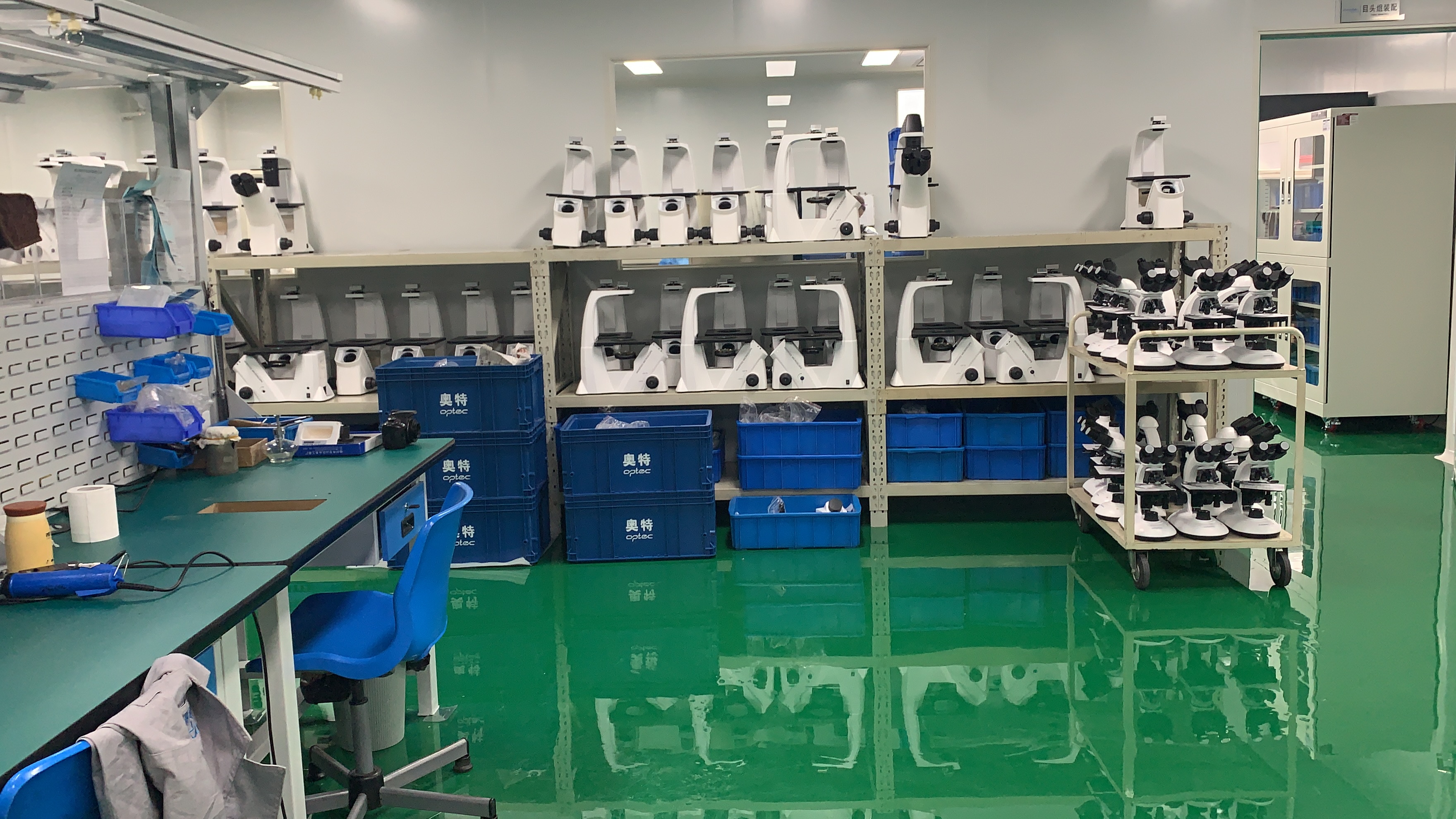
A configuration is only useful if it can be delivered, documented and repeated. Chongqing Scope Instrument Co., Ltd. operates a 5,000 m² facility with more than 100 employees, including an R&D team of 20 engineers, and reports an annual output of 30,000 units, with approximately 50% of production exported to Europe, Southeast Asia, the Middle East, South America and Africa. The company provides OEM production services and customization that includes logo customization: after technical verification, the logo can be printed on the machine, or a custom logo label can be produced and attached.
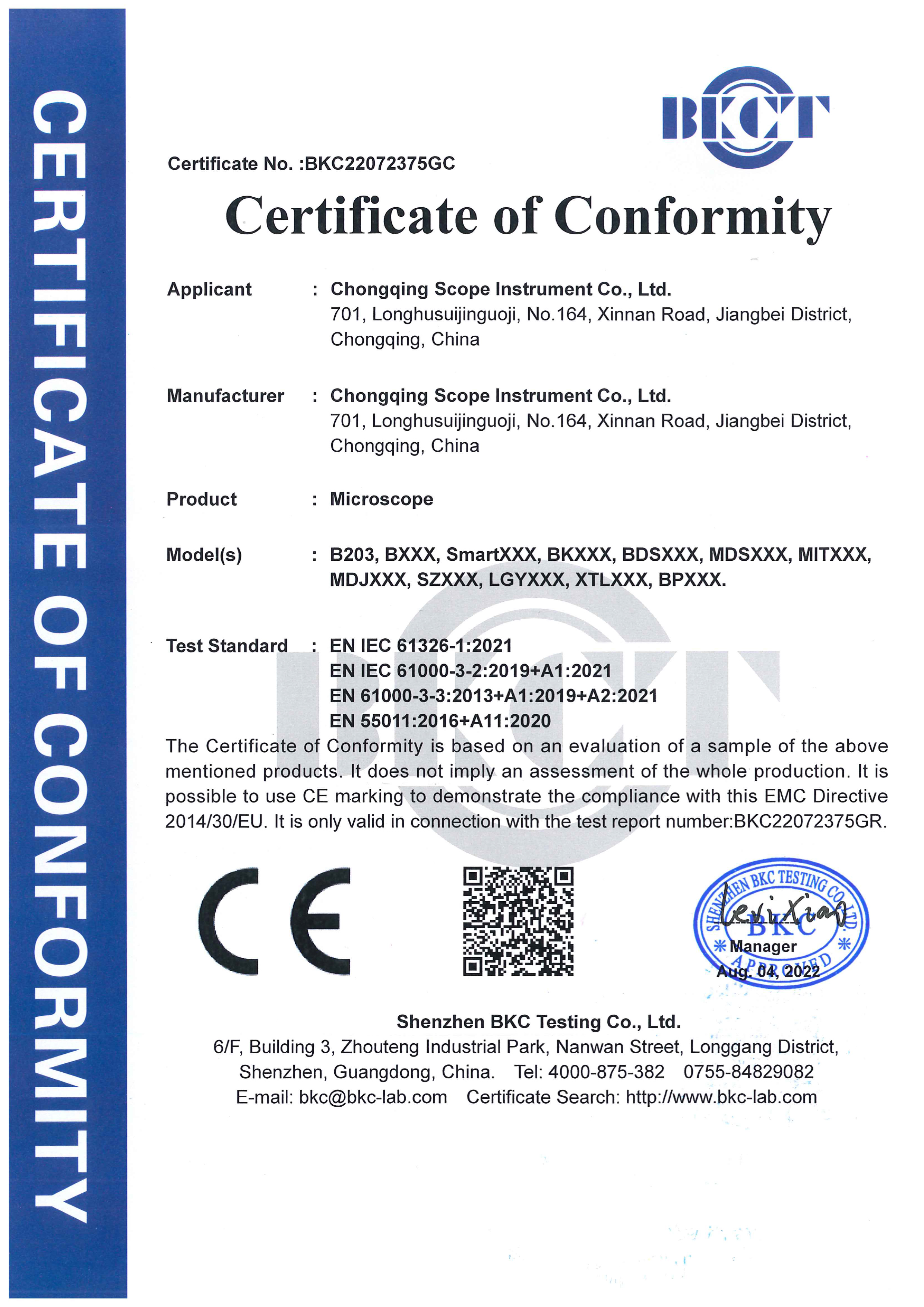
For export, certificates including ISO9001, ISO14001, ISO45001, ISO13485, CE and RoHS can be provided. Where a laboratory needs to verify compliance before purchase, the specific certificate numbers are the place to start. CE certificate BKC22072376GC, issued against EN 61010-1:2010+A1:2019 and valid until 1 August 2032, lists the B203, BXXX, BKXXX, BDSXXX, MDSXXX, MITXXX, MDJXXX, SZXXX, LGYXXX, XTLXXX and BPXXX families in its scope. CE certificate BKC22072375GC, valid until 3 August 2032, covers the electromagnetic compatibility standards EN IEC 61326-1:2021, EN IEC 61000-3-2:2019+A1:2021, EN 61000-3-3:2013+A1:2019+A2:2021 and EN 55011:2016+A11:2020 across the same family scope. RoHS certificate BKC-191103261C, valid until 18 November 2029, applies to scope B203 against the IEC 62321 series of test standards. Buyers should confirm that the certificate scope includes the exact model being ordered, and request the certificate document rather than a summary.
Reported commercial terms include a minimum order quantity of 1 unit, delivery terms of EXW, FOB, CFR, CIF or DAP, payment by T/T, acceptance criteria based on factory inspection, and a 15-day lead time, with a 1-year warranty and online support.
FAQ
Is the BDS400 or BDS500 covered by CE documentation for EU laboratories?
Chongqing Scope's CE certificate BKC22072375GC, valid until 3 August 2032, lists the BDSXXX, BKXXX, MDSXXX, MITXXX, MDJXXX, SZXXX, LGYXXX, XTLXXX, BPXXX, B203 and BXXX families within its scope, and was issued against the electromagnetic compatibility standards EN IEC 61326-1:2021, EN IEC 61000-3-2:2019+A1:2021, EN 61000-3-3:2013+A1:2019+A2:2021 and EN 55011:2016+A11:2020. A second CE certificate, BKC22072376GC, valid until 1 August 2032, was issued against EN 61010-1:2010+A1:2019 for the same family scope. The company states that ISO9001, ISO14001, ISO45001, ISO13485, CE and RoHS certificates can be provided for export. Because scope wording differs between certificates, buyers should confirm that the exact model ordered appears in the scope and request the certificate document itself rather than a summary.
Can the phase contrast configuration be customized, and can it carry our own brand?
Yes. Chongqing Scope provides OEM production services and customization that includes logo customization: once technical verification is complete, the logo can be printed on the machine, or a custom logo label can be produced and attached. The company reports a 5,000 m² facility with more than 100 employees, including an R&D team of 20 engineers, and an annual output of 30,000 units. The optical configuration — platform, objective set, condenser type and illumination — is selected per order, so branding requirements and specification requirements can be handled in the same technical discussion.
What are the minimum order quantity, delivery and payment terms?
The reported commercial terms are a minimum order quantity of 1 unit; delivery terms of EXW, FOB, CFR, CIF or DAP; payment by T/T; and acceptance criteria based on factory inspection.
Can we evaluate a single unit before committing to a larger order?
The stated minimum order quantity is 1 unit, which allows a laboratory or distributor to evaluate one configured instrument against its own specimens before scaling up. Configuration confirmation for the intended specimen type, and sample arrangements, are handled case by case.
How long is the lead time, and what support comes with the microscope?
The reported lead time is 15 days, and the microscopes are supplied with a 1-year warranty and online support. If you are specifying a phase contrast configuration for live-cell or tissue work, include the specimen type, vessel format and required magnification range in your inquiry so that the objective set, annular spot coverage and condenser type can be confirmed together. You can download the product brochure (PDF), and inquiries can be sent to claire@cqscope.com or WhatsApp +86 15223521781.
Conclusion
Low contrast in live-cell imaging is a diagnostic question before it is a purchasing decision. The specimen geometry decides whether an inverted or upright platform is appropriate; the annulus-to-objective relationship decides whether phase contrast works at all; and the objective-to-mode match decides whether the configuration stays stable across a working day. Laboratories that address these three points often find that the instrument they already own was never the problem — and those that genuinely need a phase contrast platform can now specify it precisely.
For vessel-based live-cell work, BDS500 with its 4-hole rotating disc phase contrast condenser covers 4X to 40X with matched annular positions. For thicker preparations that demand working distance, BDS400 pairs LWD Infinity Phase Contrast Objectives at 10X, 20X and 40X with a long working distance N.A. 0.3 condenser. For fixed specimens and prepared slides, BK5000-PH delivers upright phase contrast from 4X to 100X. All three are supported by OEM production, logo customization, documented export certificates, a 15-day reported lead time, and a 1-year warranty with online support.
Next step. Send your specimen type, vessel format and required magnification range to Chongqing Scope and the configuration — platform, objectives, condenser and annular spot coverage — can be quoted together. A single unit can be ordered for evaluation (MOQ 1 unit).
Email: claire@cqscope.com | Tel / WhatsApp: +86 15223521781 | Website: www.cqscopelab.com | Address: 701, Longhushuijinguoji, No.164, Xinnan Road, Yubei District, Chongqing, China
Full product brochure: download the Chongqing Scope brochure.
Have Questions or Need More Details?
Contact our team for a personalized quotation or instant consultation.
Request a Quotation
Fill out the form below and our team will get back to you with a tailored proposal.
WhatsApp Direct Chat
Prefer to chat in real-time? Message us on WhatsApp for instant assistance & quick answers.
- Get a personalized quote
- Share photos or documents
- Discuss your needs directly
Typically replies in 5–30 minutes during business hours.