
Cryoablation vs. Gamma Knife vs. S-IRE: Choosing a Minimally Invasive Cancer Treatment by Metastatic Site
Minimally invasive cancer treatment for metastatic disease is not one decision. It is a series of site-by-site decisions, made with an interventional radiologist, a radiation oncologist, a medical oncologist, and often a pathologist in the same room. Cryoablation, Gamma Knife stereotactic radiotherapy, and single-needle bipolar irreversible electroporation (S-IRE, delivered on the NanoKnife platform) are three of the tools that sit at the centre of that sequence — and the published case record at Guangzhou Fuda Cancer Hospital shows them used inside one multidisciplinary (MDT) treatment model rather than as competing versions of the same idea.
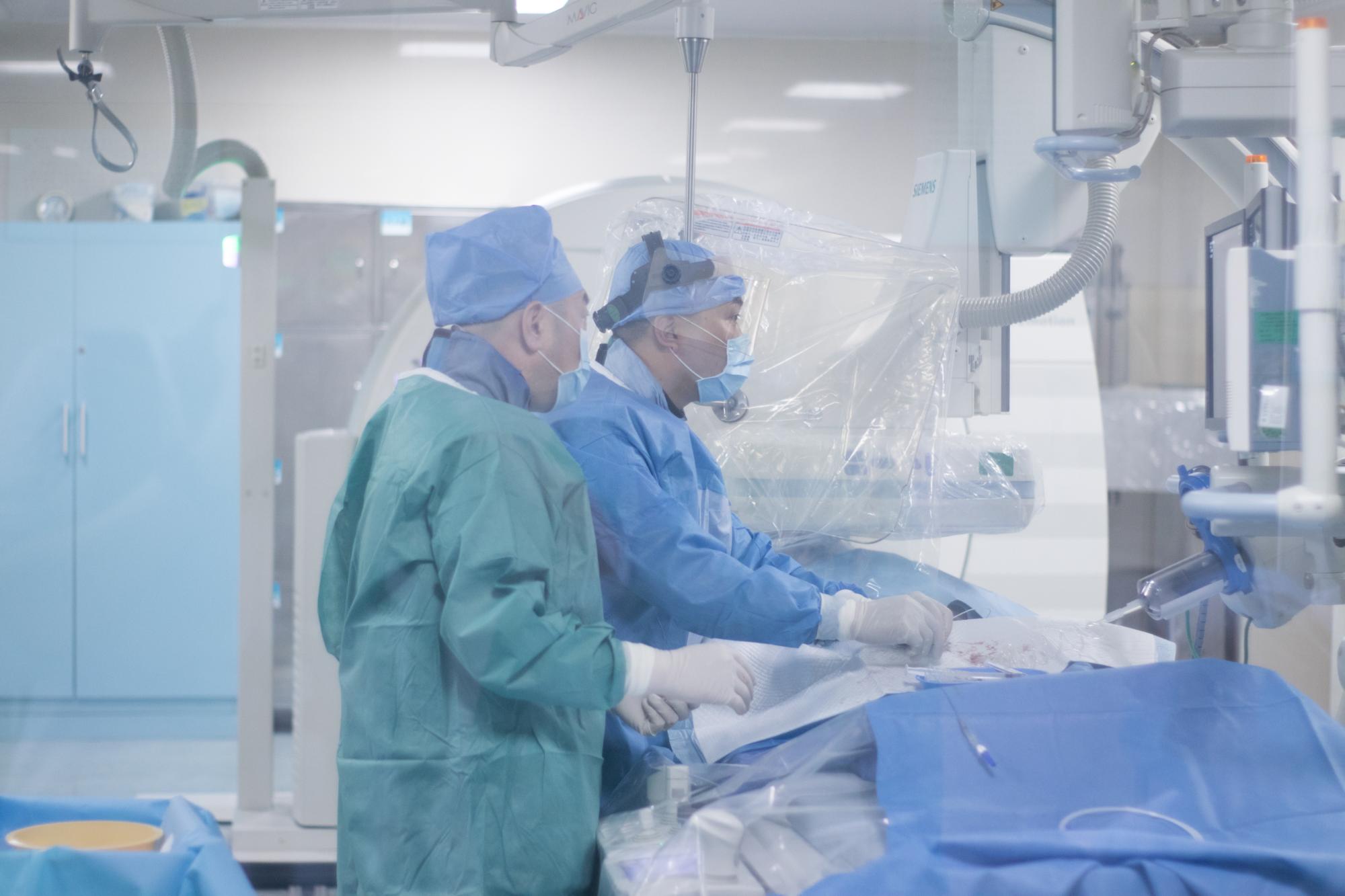
Cover: an image-guided interventional suite — the setting where percutaneous cryoablation and single-needle S-IRE are performed.
No single modality in this comparison is documented as universally superior. In the case reports reviewed below, the metastatic site decided the tool: an accessible chest-wall deposit in metastatic triple-negative breast cancer was treated with cryoablation combined with a biopsy; meningeal metastasis in the same patient was treated with Gamma Knife inside the same MDT plan; and a recurrent pancreatic lesion of approximately 7 cm after Whipple surgery, encasing the celiac trunk and the superior mesenteric artery, was treated with percutaneous S-IRE under CT and ultrasound guidance, with the patient under local anesthesia. The remainder of this guide sets out the criteria behind those choices.
Problem Definition: Why 'Which One Is Best?' Misses the Point
The most common question from patients and referring physicians is a ranking question: which of these three is the strongest option for metastatic cancer? That framing produces bad answers, because the three techniques do not compete on the same axis. They differ in how they destroy tissue, whether they require a percutaneous track, whether a tissue sample can be taken at the same time, what anesthesia they need, and how easily they can be combined with systemic therapy.
A second problem is sequencing. In metastatic disease, a patient rarely has one lesion in one organ. The same person may present with a chest-wall deposit, a meningeal finding, and a dominant visceral mass, each with different accessibility and different urgency. Deciding once, for the whole patient, tends to produce a plan that is a good fit for one site and a poor fit for the others.
The practical reframing is therefore: for this lesion, at this site, in this treatment sequence, with this patient's tolerance — which tool does the least harm for the most local control? That question can be answered with documented criteria. A ranking question cannot.
What this comparison is not. It is not a claim that one technique should replace the others, and it is not a survival comparison. The evidence used here is limited to documented case reports and to published criteria such as lesion location, biopsy requirement, anesthesia and recovery, and integration with systemic therapy. Where the published record does not state something, this guide says so rather than filling the gap.
Industry Background: Where These Three Options Sit in 2026
Ablation and stereotactic techniques have moved from last-resort palliation toward planned, organ-directed treatment. The commercial signal is visible: the global minimally invasive surgery market is projected to reach USD 199.30 billion by 2030, according to MarketsandMarkets (Minimally Invasive Surgery Market Report 2025–2030). Regionally, Asia Pacific accounted for a 37.6% revenue share of the cancer treatment facilities market in 2025, according to Grand View Research — a regional supply base that now supports a high volume of image-guided oncology work.
Regulatory maturity matters for patients comparing providers. Cryosurgery for cancer treatment was approved by China's State Drug Administration (SDA) in 1999, meaning the technique has a long regulated history rather than a recent experimental status. Clinical recognition has followed: research from Fuda Cancer Hospital on cryoablation for lung nodules was cited in the 2024 expert consensus of the American Association for Thoracic Surgery (AATS).
Institutional depth is the other half of the background. Guangzhou Fuda Cancer Hospital was founded in 2003 and is an oncology-specialised hospital operating the Tianhe Campus and the Haizhu Campus, with a total floor area of over 30,000 m² and 400 open beds. It is the first oncology-specialised hospital in Guangdong Province accredited by Joint Commission International (JCI), and in 2010 it was designated as one of the first batch of National Key Clinical Cancer Speciality Centres (Oncology). Its research and clinical teams focus on cryoablation, irreversible electroporation (NanoKnife), interventional oncology, and iodine seed therapy — which is precisely why the same institution documents all three modalities discussed here. Patients have been received from more than 130 countries and regions, with about 60% arriving from Southeast Asia, the Middle East, Europe and North America.
Detailed Solution: What Each Modality Actually Does
The three techniques are best understood by their energy source and their access route. That is also what determines which metastatic sites they suit.
Cryoablation: thermal ablation through controlled freezing
Cryoablation is a local ablation technique that destroys tumour tissue by freezing it through a probe placed into the lesion. In the documented metastatic breast cancer case reviewed later in this article, cryoablation was performed on an accessible chest-wall lesion and was delivered together with a biopsy in the same procedure. Guangzhou Fuda Cancer Hospital reports having completed over 10,000 cases of cryosurgery across more than 30 cancer types, covering lung, liver and soft-tissue work among others. The general trade-off is accessibility: the lesion has to be reachable with a probe, and the surrounding structures have to tolerate a freezing zone.
Gamma Knife: stereotactic radiotherapy without a percutaneous track
Gamma Knife is a stereotactic radiotherapy platform rather than a needle-based ablation tool. It requires no incision and no probe track, which makes it relevant for lesions that are difficult or unsafe to puncture — the documented example being meningeal metastasis in the metastatic breast cancer case, planned and executed through Gamma Knife planning and execution records within an MDT framework. The trade-off is that it delivers radiation rather than immediate tissue destruction, and it does not produce a pathology specimen in itself.
S-IRE: single-needle bipolar irreversible electroporation (NanoKnife)
S-IRE is a non-thermal ablation technique that uses an electric field rather than heat or cold, delivered here through a single needle. That access route matters in the documented pancreatic case, where the lesion encased the celiac trunk and the superior mesenteric artery: the procedure was performed percutaneously under combined CT and ultrasound (dual-image) guidance, with the patient under local anesthesia, and the same session included concurrent pathology sampling and a celiac plexus block. Because the approach is percutaneous, tissue can be obtained for pathology at the same time — a capability that distinguishes it from pure radiotherapy approaches.
Four Criteria That Decide the Choice by Site
1. Lesion location and accessibility
Accessibility is the first filter, and it is anatomical rather than statistical. An accessible chest-wall nodule can be approached with a probe. A meningeal lesion is better addressed with a non-invasive stereotactic approach. A deep pancreatic lesion wrapped around major vessels is the case where single-needle electric-field ablation was documented, using real-time CT and ultrasound guidance rather than open access. Questions worth asking a treating team are: can the lesion be reached without crossing critical structures, and what image guidance will confirm probe placement in real time?
2. Whether a concurrent biopsy is needed
In metastatic disease, a new lesion is often the moment when tissue confirmation and molecular profiling become clinically necessary. This is a decision criterion in its own right, not an afterthought. The documented pancreatic S-IRE procedure included concurrent pathology sampling in the same session, and the documented breast cancer pathway lists biopsy combined with cryoablation as a single step. In both instances, the ablation route doubled as a diagnostic route. Gamma Knife does not provide that function, so a separate biopsy is planned when tissue is required.
3. Anesthesia, procedure setting, and recovery
Anesthesia requirements shape who can be treated and how quickly they return to daily life. In the documented S-IRE pancreatic case, the procedure was performed under local anesthesia, took place in a single session, and the patient ambulated the next day with short postoperative inpatient observation. The report also records satisfactory pain control, supported by a celiac plexus block performed during the same procedure. For Gamma Knife, the relevant burden is planning and delivery rather than recovery from a puncture. These differences are often the deciding factor for frail or previously operated patients — including those who have already undergone major surgery such as a Whipple resection.
4. Integration with systemic therapy
Local control in metastatic disease is rarely an alternative to systemic treatment; it sits inside it. The documented breast cancer pathway combined local ablation and stereotactic radiotherapy with molecular marker-guided systemic therapy, dosed as ADC plus immuno/targeted therapy, with an imaging recheck after two cycles. The pancreatic case was planned and delivered through a multidisciplinary framework with post-procedure imaging review and follow-up recommendations for continuing monitoring. When evaluating any programme, ask how the ablation or radiosurgery date is coordinated with the systemic cycle — the sequencing decision is as consequential as the modality decision.
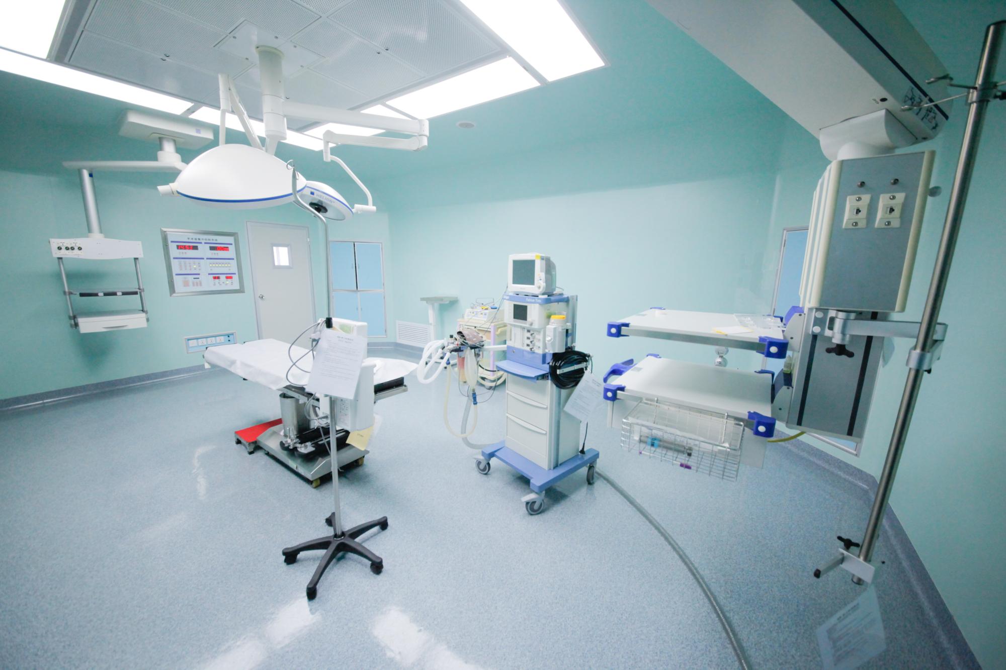
Procedure room: anesthesia planning, image guidance and concurrent tissue sampling are decided together before the session, not during it.
Step-by-Step: How a Site-Specific Plan Is Actually Built
Across the documented cases, the same pathway repeats. It is named Consultation → MDT → Treatment → Follow-up, and its five phases are: appointment and initial visit; examination and assessment; MDT decision; treatment implementation; and discharge and follow-up.
- Appointment and initial visit. The entry point is an online or phone appointment followed by an outpatient assessment. The patient or referring physician is expected to provide a complete medical history and test records.
- Examination and assessment. Imaging and molecular tests are performed, together with pathology review. This is the stage that determines whether a lesion is accessible, whether tissue is needed, and what anesthesia is realistic.
- MDT decision. The multidisciplinary team formulates the plan, including the personalized treatment plan and procedure scheduling. Local ablation, stereotactic radiotherapy and systemic therapy are sequenced at this point rather than chosen separately.
- Treatment implementation. Procedures are carried out with image guidance. In the documented pancreatic case, this meant preoperative MDT review, image-guided puncture, single-needle energy delivery, concurrent biopsy and celiac plexus block, in one session under local anesthesia.
- Discharge and follow-up. Stage outputs include postoperative management and a follow-up schedule. Reviews are conducted through MDT and follow-up for efficacy assessment, and the revision policy allows the plan to be adjusted on the basis of imaging, pathology or treatment response, with the MDT reconvened when a change is indicated.
That last point is the difference between an ablation procedure and a treatment programme. A one-off procedure produces one imaging comparison. A programme produces imaging comparisons, pathology results, recovery records and follow-up recommendations — and changes course when the imaging says so.
Use Cases: Two Documented Metastatic Scenarios
Case 1 — Recurrent pancreatic cancer encasing the celiac trunk and superior mesenteric artery
The lesion was a recurrent pancreatic tumour of approximately 7 cm after Whipple surgery, encasing the celiac trunk and the superior mesenteric artery. Treatment was percutaneous S-IRE using a single needle under combined CT and ultrasound guidance, with the patient under local anesthesia. The methodology recorded for the case included multidisciplinary team evaluation, real-time dual-image guidance, single-needle electric-field ablation, concurrent pathology sampling and analgesia control.
The reported execution sequence was: preoperative multidisciplinary team review, image-guided puncture, single-needle energy delivery, concurrent biopsy and celiac plexus block, and postoperative imaging review with short rehabilitation. MDT discussion and preparation took days and the procedure was completed in a single session followed by short postoperative inpatient observation; the exact duration was not stated in the report. Quantitative results reported that the patient ambulated the next day, and CT imaging showed evident necrosis and shrinkage of the lesion — no numeric values were provided. Qualitative results reported good recovery, satisfactory pain control and positive family feedback. Proof assets attached to the official report include pre-treatment CT images dated August 5 and post-treatment CT images dated August 14, together with procedure photographs. The case was published as an anonymized hospital case report in the patient story section.
Case 2 — Metastatic triple-negative breast cancer with meningeal involvement
This case used a combination rather than a single modality: chest-wall cryoablation plus Gamma Knife for meningeal metastasis, delivered within one MDT plan. The methodology was an MDT individualized plan integrating image-guided ablation, stereotactic radiotherapy and molecular marker-guided systemic therapy.
The execution sequence was: admission assessment, MDT, complication management, biopsy combined with cryoablation, Gamma Knife, ADC plus immuno/targeted dosing, and imaging recheck after two cycles. The project duration was about two months, covering local interventions and two systemic treatment cycles with imaging reassessment. Deliverables included inpatient records, biopsy and ablation procedural records, Gamma Knife planning and execution records, imaging comparisons and follow-up recommendations.
Quantitative results showed approximately 50% radiologic reduction of overall tumour lesions within about two months. Qualitative improvements included reduced pain, reduced dyspnoea and improved exercise tolerance, with the patient progressing from wheelchair dependence to walking longer distances. Non-measurable benefits reported were improvement in quality of life and functional mobility. The case is anonymized; the published page includes pre- and post-treatment imaging comparisons, while original imaging and pathology reports require provision by the hospital for medical proof.
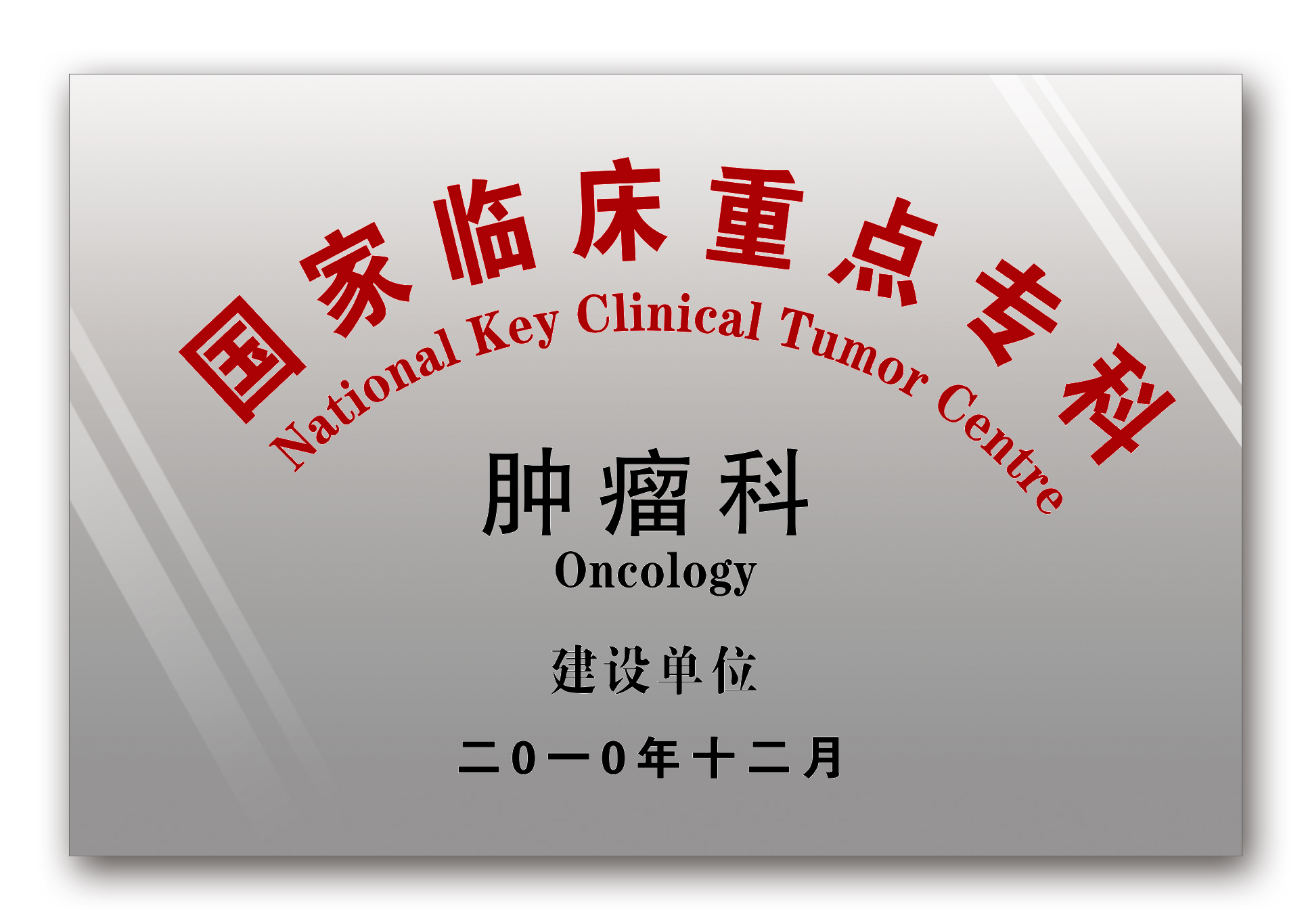
Proof: national key clinical specialty (oncology) recognition — the institutional layer behind the case reports.
Site-by-Site Comparison: What the Case Records Actually Show
The table below is deliberately restricted to what the two documented cases state. Empty cells mean the published report does not specify the detail; they are not evidence that a modality cannot do it.
| Comparison criterion | Cryoablation | Gamma Knife | S-IRE (single-needle bipolar NanoKnife) |
|---|---|---|---|
| Lesion site documented in these cases | Chest-wall deposit (metastatic triple-negative breast cancer) | Meningeal metastasis (same MDT plan) | Recurrent pancreatic lesion approx. 7 cm post-Whipple, encasing celiac trunk and superior mesenteric artery |
| Energy / approach | Thermal ablation (controlled freezing) with a probe placed in the lesion | Stereotactic radiotherapy; no percutaneous track | Non-thermal electric-field ablation delivered through a single needle, percutaneously |
| Image guidance documented | Not specified in the published case summary | Gamma Knife planning and execution records | Real-time dual-image guidance (CT + ultrasound) |
| Concurrent tissue sampling | Yes — biopsy combined with cryoablation in the documented sequence | Not documented in this case; a separate biopsy is required if tissue is needed | Yes — concurrent pathology sampling during the same procedure |
| Anesthesia / pain management documented | Not specified in the published case summary | Not specified; non-invasive delivery, no puncture | Local anesthesia with analgesia control; concurrent celiac plexus block |
| Early recovery documented | Not specified in the published case summary | Not specified in the published case summary | Ambulatory the next day; short postoperative inpatient observation after a single session |
| Integration with systemic therapy | Yes — part of an MDT plan that also included ADC plus immuno/targeted dosing | Yes — planned within the same MDT framework as the systemic regimen | Planned and delivered through a multidisciplinary framework, with post-procedure imaging review and follow-up recommendations for continuing monitoring |
| Reported outcome in the documented case | Approx. 50% radiologic reduction of overall tumour lesions within about two months (plan-level result) | Included in the same two-month plan and imaging reassessment | CT showed evident necrosis and shrinkage of the lesion; no numeric values provided |
Table 1. Site-specific trade-offs as documented in two anonymized case reports — not a universal ranking of modalities.
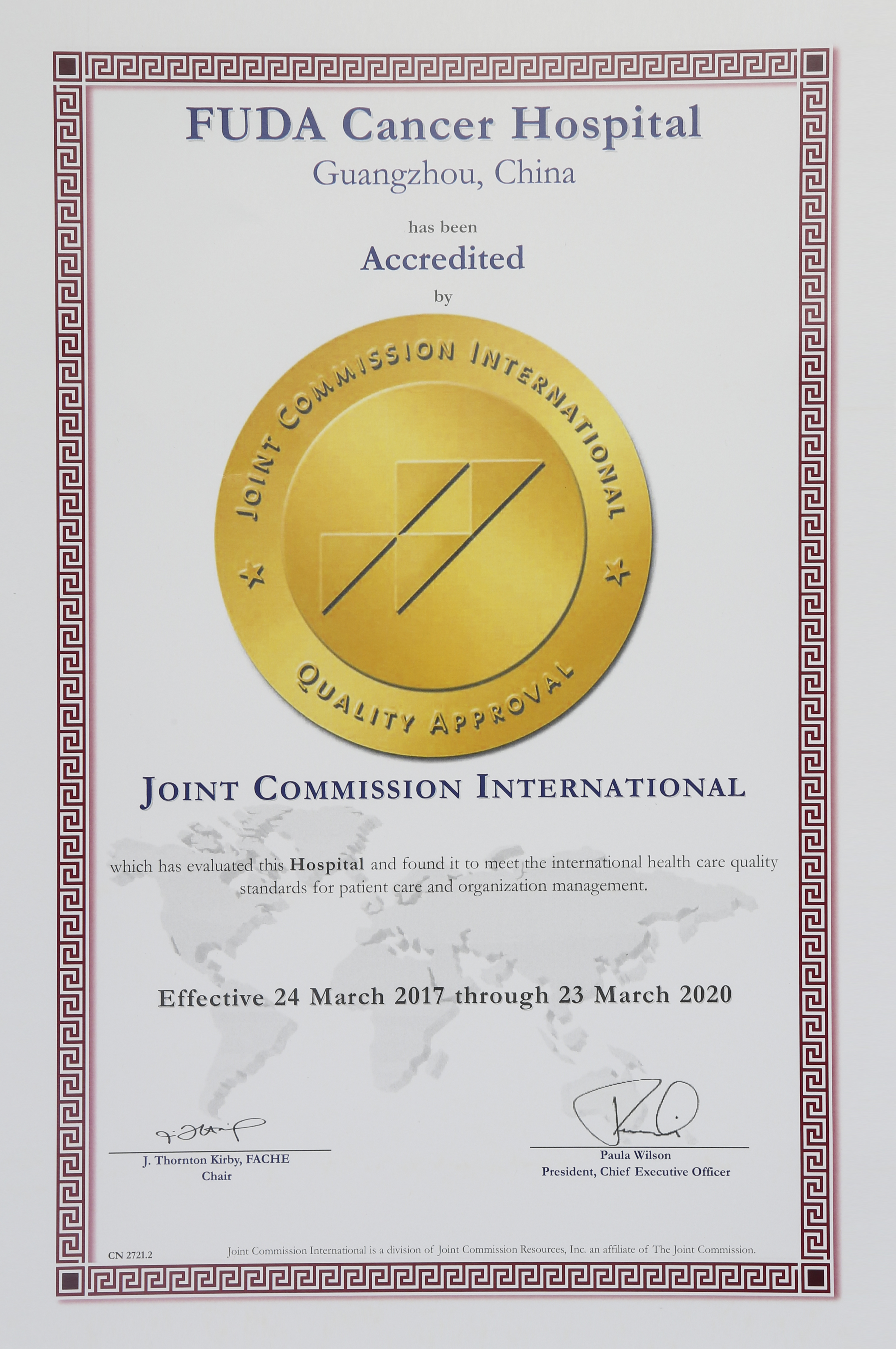
Compliance layer: JCI accreditation, held with re-evaluation documented, is the institutional quality check behind the technique approvals.
Frequently Asked Questions
Is cryoablation a formally approved cancer treatment, and how is hospital quality verified?
These are two separate checks. Cryosurgery for cancer treatment was approved by China's State Drug Administration (SDA) in 1999, which addresses the regulatory status of the technique in the Chinese market. Institutional quality is verified differently: JCI accreditation reflects compliance with international patient safety and quality standards, while national key clinical specialty designations reflect clinical review. Guangzhou Fuda Cancer Hospital is the first oncology-specialised hospital in Guangdong Province accredited by Joint Commission International (JCI), with re-evaluation documented, and it was designated in 2010 as one of the first batch of National Key Clinical Cancer Speciality Centres (Oncology), later accredited as a National Key Clinical Specialty (Oncology) in 2018. For patients comparing providers, the practical rule is to request both: the technique's approval status and the hospital's accreditation status.
Can one minimally invasive technique treat a metastasis in any location?
No — in the documented cases, location changed the tool. A chest-wall deposit in metastatic triple-negative breast cancer was treated with cryoablation combined with a biopsy. Meningeal metastasis in the same patient was treated with Gamma Knife inside the same MDT plan. A recurrent pancreatic lesion encasing the celiac trunk and superior mesenteric artery was treated with percutaneous S-IRE using a single needle under CT and ultrasound guidance, with the patient under local anesthesia. Capability therefore sits at the level of the treatment programme, not the device: the relevant question is whether the centre plans image guidance, anesthesia, tissue sampling and systemic therapy around the specific site.
If the plan is personalized, how is a case-specific estimate determined?
The documented pathway runs Consultation → MDT → Treatment → Follow-up: online or phone appointment, outpatient assessment, imaging and molecular tests, then an MDT decision before treatment implementation. Because the personalized treatment plan and the procedure schedule are outputs of the assessment phase, a case-specific estimate follows record review and imaging rather than preceding it. Patients and referring physicians are therefore asked first for the complete medical history, imaging, pathology or molecular results and prior treatment records. Sending those records to the hospital's international patient coordinator is the standard way to begin the review: appointment@fudahospital.com or WhatsApp +86 189-2215-3602.
What documentation accompanies treatment, and can it be verified?
Documented deliverables include inpatient records, biopsy and ablation procedural records, Gamma Knife planning and execution records, imaging comparisons and follow-up recommendations. Other case records list a treatment plan document, operative and interventional records, imaging and tumour-marker follow-up records, procedure records, a discharge and follow-up plan, comparative imaging at admission versus after two treatment sessions, MDT notes and nursing records. Published cases are anonymized, and original imaging and pathology reports can be requested from the hospital for medical proof. For a referring physician, this package functions like a validation sample: it shows what was planned, what was performed and what changed on imaging.
How long does preparation take, and what is the first step?
Preparation time is case-specific. In the reported pancreatic S-IRE case, multidisciplinary team discussion and preparation took days, and the procedure was completed in a single session followed by short postoperative inpatient observation; the exact duration was not stated in the report. In the reported metastatic breast cancer case, the overall treatment period was about two months, covering local interventions plus two systemic treatment cycles with an imaging reassessment. The first step is an online or phone appointment, followed by outpatient assessment, imaging and molecular tests, and then the MDT decision that produces the personalized plan.
Conclusion: Choose by Site, Then Commit to the Programme
The three modalities reviewed here answer different anatomical questions. Cryoablation is documented for an accessible chest-wall lesion, delivered together with biopsy. Gamma Knife is documented for meningeal metastasis, delivered without a puncture track. Single-needle S-IRE is documented for a recurrent pancreatic lesion encasing major vessels, delivered percutaneously under CT and ultrasound guidance with local anesthesia and concurrent tissue sampling. None of these is a general answer; each is a site-specific answer.
What should carry more weight than the modality name is the structure around it. In both documented cases, the result came from a sequence — MDT assessment, imaging and molecular testing, a personalized combined plan, and follow-up with the option to revise the plan when imaging, pathology or response indicates a change. That structure, not the device label, is what makes a minimally invasive cancer treatment plan durable over months of metastatic disease management.
Next Step: Case Review and Sample Materials
Guangzhou Fuda Cancer Hospital reviews cases for patients and referring physicians, including those already treated elsewhere. For a case review, send the medical history, imaging, pathology or molecular results and prior treatment records to the international patient coordinator: appointment@fudahospital.com or WhatsApp +86 189-2215-3602. You can also download the hospital brochure for full treatment information: Fuda Cancer Hospital brochure (PDF). Hospital website: www.fudahospital.com. Address: No. 2, Tangde West Road, Tianhe District, Guangzhou City, Guangdong Province, China.

CTA: the consultation pathway begins with an online or phone appointment and an outpatient assessment.
Have Questions or Need More Details?
Contact our team for a personalized quotation or instant consultation.
Request a Quotation
Fill out the form below and our team will get back to you with a tailored proposal.
WhatsApp Direct Chat
Prefer to chat in real-time? Message us on WhatsApp for instant assistance & quick answers.
- Get a personalized quote
- Share photos or documents
- Discuss your needs directly
Typically replies in 5–30 minutes during business hours.