
Ewing Sarcoma Lung Metastasis at 17 cm: A Non-Surgical Interventional Scenario
Ewing sarcoma that has spread to the lung can produce a metastasis too large, too central, or too close to vital structures to be removed safely. The documented case summarised here is one of those situations. A young patient from Lebanon with Ewing sarcoma pulmonary metastasis had a lesion sitting adjacent to the pericardium and major vessels, and the tumor grew from 8 cm to nearly 17 cm. Surgical resection was therefore not a workable plan. Treatment was carried out at Fuda Cancer Hospital in Guangzhou, China, using superselective interventional embolization-chemotherapy in staged sessions, followed by combined targeted drug management and periodic CT follow-up. In the hospital's case record, the tumor decreased from nearly 17 cm to approximately 5 cm.
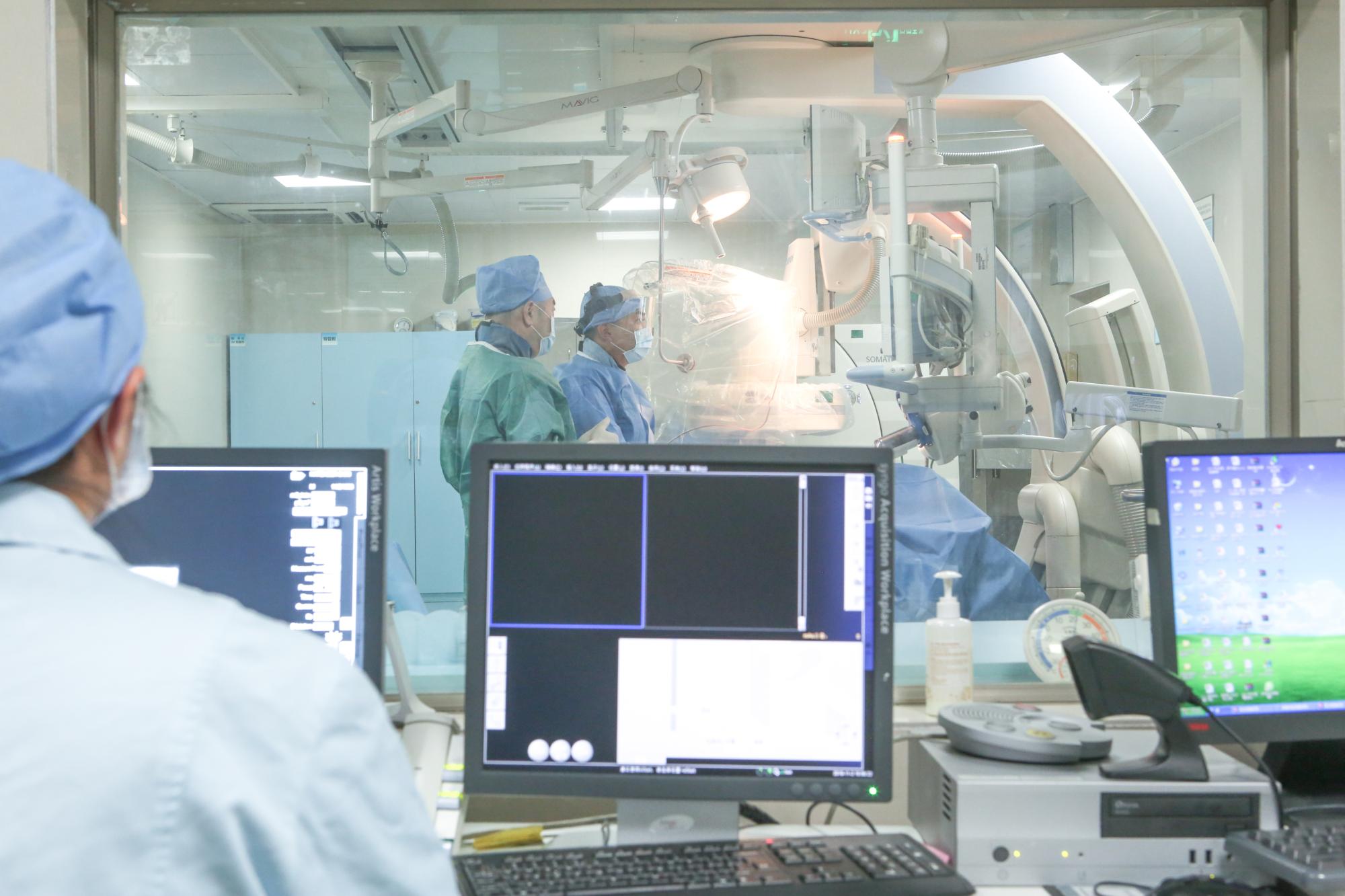
Problem Definition: Why a 17 cm Lung Metastasis Could Not Be Operated On
Three specific features pushed the treatment decision away from surgery and towards an image-guided, interventional approach.
- Anatomical position. The metastatic lesion was adjacent to the pericardium and major vessels. In that location, the margin between tumor and vital structures is measured in millimetres, and the risk of an operation is not confined to the tumor itself.
- Growth trajectory. The lesion had grown from 8 cm to nearly 17 cm. A mass of that size and rate of change removes the option of a straightforward, single-step resection.
- Pathological complexity. In Ewing sarcoma lung metastasis, the pathological differentiation is complex, and the morphology can be confused with small cell lung cancer. Confirming the diagnosis through pathology review is a prerequisite, because it determines which treatment combinations are appropriate.
The practical question for families in this position is rarely whether surgery would be ideal. It is what can be done when surgery is not available as a safe option, and how the result can be measured and documented over time.
Industry Background: Where Interventional Oncology Fits
Interventional oncology treats tumors through image-guided, minimally invasive procedures rather than open resection. Catheters, probes and needles are navigated into or beside a lesion under imaging guidance, so that treatment acts locally while systemic therapy addresses the wider disease. For tumors that are unresectable, locally advanced or recurrent, this branch of oncology provides treatment pathways that do not depend on removing the tumor in one operation.
The wider setting explains why this type of care has expanded. China's hospital services market was estimated at USD 614.82 billion in 2024, with specialised private hospitals described as a significant growth driver (Market Research Future). China's healthcare expenditure for cancer treatment reached 221.4 billion RMB, accounting for 5.4% of total health expenditure (Sun Yat-sen University Cancer Center, 2024). On the technology side, the global cryoablation devices market is projected to grow from USD 614.3 million in 2026 to USD 1,036.7 million by 2030, with hospitals as the largest end-user segment at 54.4% (Grand View Research, 2026).
Regulatory history matters as well. Irreversible electroporation (NanoKnife) was approved for clinical application in China in June 2015, and Fuda Cancer Hospital reports being the first institution to introduce the therapy in the country. For international patients, however, technology is only half of the picture. Cross-border care also depends on language support, coordinated scheduling and a follow-up structure that continues after the patient returns home.
Detailed Solution: 3C+P and Superselective Interventional Embolization-Chemotherapy
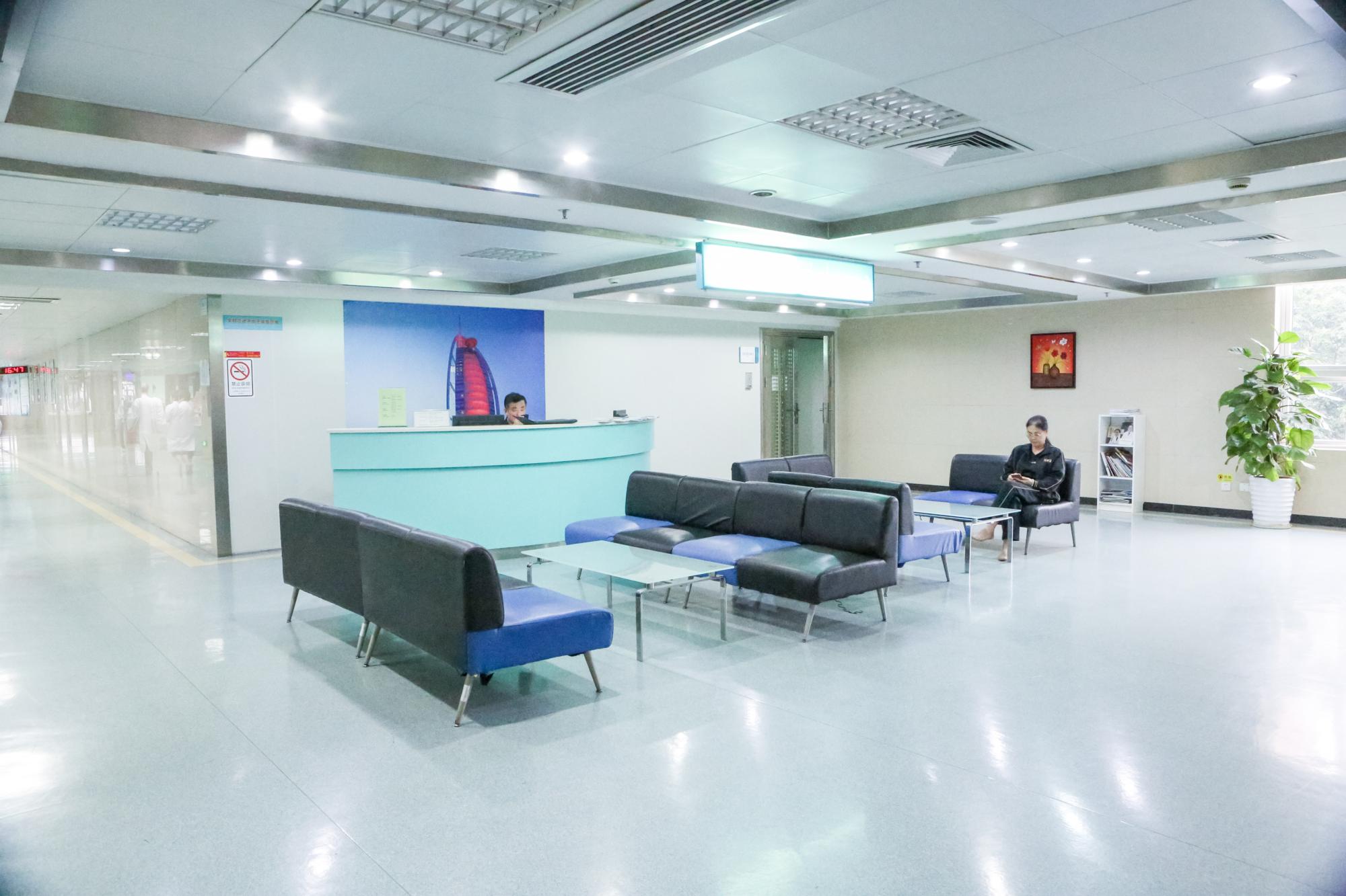
Guangzhou Fuda Cancer Hospital (Fuda Cancer Hospital) is an oncology-specialised hospital in Guangzhou, China, operating the Tianhe and Haizhu campuses with a total floor area of more than 30,000 m2, 400 open beds and 45 VIP rooms. It is accredited by Joint Commission International (JCI) and is described by the hospital as the first oncology-specialised hospital in Guangdong Province to hold that accreditation. It was designated a National Key Clinical Specialty (Oncology) in 2018 and named a High-level Key Clinical Cancer Speciality Centre of Guangdong Province (Oncology) in 2019. Patients from more than 130 countries and regions have been treated there, with about 60% coming from Southeast Asia, the Middle East, Europe and North America.
The clinical model used across the hospital is the 3C+P Personalized Comprehensive Oncology Solution, which combines:
- Cryo-Irreversible Electroporation Ablation (CIA) — cryoablation performed with image-guided cryo probes, and irreversible electroporation (NanoKnife) offered for inoperable tumors as a non-surgical treatment option.
- Cancer Vascular Intervention (CVI) — vascular interventional oncology, including superselective embolization with local high-concentration chemotherapy.
- Combined Immunotherapy for Cancer (CIC) — combined immunotherapy supported by molecular testing that informs targeted therapy evaluation and systemic treatment planning.
- Personalized (P) comprehensive therapy — MDT-based decisions, sequencing of local and systemic treatment, rehabilitation and scheduled follow-up.
In this patient, the component applied was the vascular interventional part of the model: superselective interventional embolization-chemotherapy. The hospital describes the methodology as MDT assessment combined with image-guided superselective embolization and local high-concentration chemotherapy, delivered over multiple staged sessions, then combined with targeted drug management and periodic CT follow-up. The supporting technology base includes 64-slice CT, 1.5T MRI, DSA, cryo probes, NanoKnife, microwave ablation, radiotherapy and chemotherapy devices, and molecular diagnostics — each of which becomes relevant or irrelevant depending on the lesion being treated.
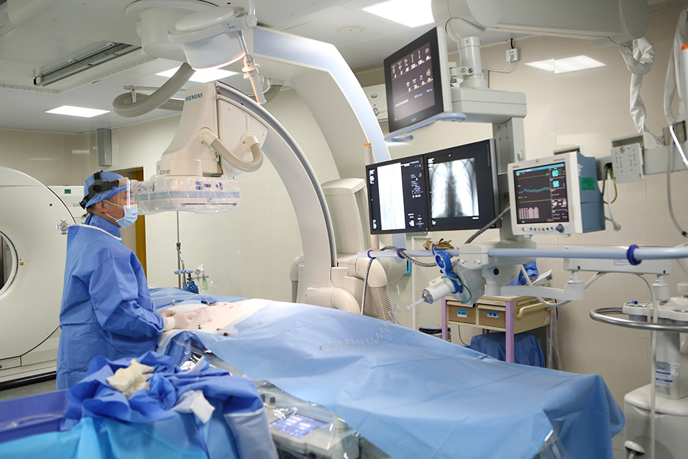
Step-by-Step: From Admission Evaluation to Long-Term Follow-Up
The general pathway at the hospital runs through outpatient evaluation, admission preparation, operative or interventional procedures, post-operative monitoring, and rehabilitation and follow-up. In this case, the documented execution steps were:
- Admission evaluation — medical history, prior treatment records and current symptoms were reviewed as the basis for treatment planning.
- Imaging and pathology review — imaging defined the size and position of the pulmonary lesion; pathology review addressed the complex differentiation of Ewing sarcoma lung metastasis.
- MDT decision — a multidisciplinary team concluded that resection was not the appropriate route and selected superselective interventional embolization-chemotherapy.
- Staged embolization-chemotherapy sessions — multiple interventional treatments delivered local high-concentration chemotherapy into the tumor-feeding vessels, with embolization, using image guidance.
- Imaging rechecks — CT follow-up between and after sessions tracked the change in lesion size, which is how the reduction from nearly 17 cm to approximately 5 cm was recorded.
- Combined targeted therapy and long-term follow-up — targeted drug management continued after the interventional phase, with periodic CT assessment and a schedule designed to continue after discharge.
The case record also notes tolerability: symptom relief, good tolerance of treatment, no significant vomiting and only mild skin dryness were reported. Tolerability matters in advanced disease, because it determines whether a staged plan can actually be completed.
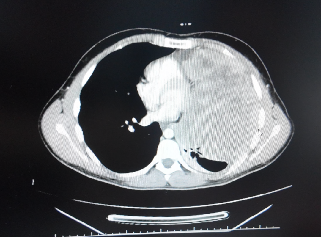
Documents Patients Can Request After Treatment
For a cross-border patient, the treatment record is not an administrative detail — it is the handover document that a physician at home will work from. The deliverables documented in this case were:
- Imaging comparison reports (before and after treatment)
- Procedure records for each interventional session
- Discharge and follow-up plan
- Nursing records
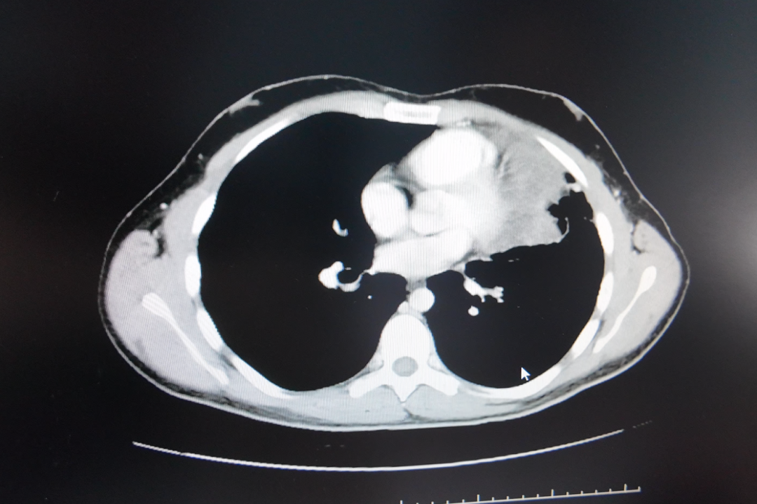
Use Cases: Non-Surgical Interventional Scenarios
Primary case: Ewing sarcoma lung metastasis, Lebanon to Guangzhou
A young student from Lebanon presented with Ewing sarcoma pulmonary metastasis that had grown from 8 cm to nearly 17 cm, with the lesion adjacent to the pericardium and major vessels. Because a surgical plan was not feasible, the treatment route was superselective interventional embolization-chemotherapy in multiple staged sessions, followed by combined targeted drug management and periodic CT follow-up. The recorded outcome was a decrease from nearly 17 cm to approximately 5 cm, with good tolerance and symptom relief. The patient's feedback in the case record expressed satisfaction with the treatment effect and the care received.
Comparable scenarios documented at the same hospital
Metastatic breast cancer, Indonesia. A patient whose prior multi-line chemotherapy and radiotherapy were ineffective, with multiple systemic metastases and severe functional decline, was managed through MDT assessment, biopsy, cryoablation for a chest wall lesion, Gamma Knife treatment for meningeal metastasis, and ADC combined immuno/targeted therapy guided by molecular markers. Reassessment within about two months recorded an approximately near-50% radiologic reduction across lesions, with the patient progressing from wheelchair dependence to walking longer distances.
Recurrent pancreatic cancer, Hong Kong. An 81-year-old patient with a recurrent lesion encasing the celiac trunk and superior mesenteric artery was judged unable to tolerate resection or general anaesthesia. The team performed percutaneous single-needle bipolar nanoknife ablation under combined CT and ultrasound guidance with local anaesthesia, together with concurrent biopsy and a celiac plexus block. The record notes that the patient ambulated the next day and that CT showed evident necrosis or shrinkage of the lesion.
Lung squamous carcinoma with brain metastases, Malaysia. Treatment combined Gamma Knife for brain metastases with interventional arterial perfusion chemotherapy for thoracic and systemic disease, together with a change of immunotherapy. Imaging showed lesion shrinkage, the tumour marker CYFRA21-1 decreased to the normal range in the record, and the patient resumed daily activities without severe adverse effects.
These are individual, anonymised case records from one hospital. They illustrate the type of clinical situation an interventional and MDT-based model is designed for; they are not a prediction that another patient will achieve the same result.
Comparison Table: 3C+P Components and Their Role in This Case
The 3C+P model is not applied as a package to every patient. MDT evaluation determines which components are used, in what order and in how many sessions.
| Component of the 3C+P model | Therapies and modules included | Role in this pulmonary metastasis case |
|---|---|---|
| Cryo-Irreversible Electroporation Ablation (CIA) | Cryoablation with image-guided cryo probes; irreversible electroporation (NanoKnife) offered for inoperable tumors as a non-surgical option | Not the primary approach used here. Relevant when a lesion can be reached percutaneously and local ablation is judged appropriate by the MDT. |
| Cancer Vascular Intervention (CVI) | Image-guided vascular interventional oncology, including superselective embolization with local high-concentration chemotherapy | The component applied in this case, delivered across multiple staged interventional sessions. |
| Combined Immunotherapy for Cancer (CIC) | Combined immunotherapy integrated with molecular testing that supports targeted therapy evaluation and systemic treatment planning | The case record describes combined targeted drug management after the interventional sessions, with periodic CT follow-up. |
| Personalized (P) comprehensive therapy | MDT evaluation, individualized treatment decisions, rehabilitation and scheduled follow-up | MDT review set the treatment sequence, and follow-up continued on a scheduled basis with imaging assessment. |
FAQ: Multidisciplinary Oncology Teams and Non-Surgical Treatment
What does a multidisciplinary oncology team actually decide?
A multidisciplinary team (MDT) brings specialists together to make one coordinated decision for a complex case, rather than a series of separate opinions. At Fuda Cancer Hospital, MDT evaluation is a module of the 3C+P Personalized Comprehensive Oncology Solution and supports multidisciplinary, individualised treatment decisions for complex cases. In practice, the MDT reviews imaging, pathology and molecular results, then decides whether the case calls for ablation, vascular intervention, systemic therapy or a combination — and in what order. This structure is supported by the hospital's accreditation and specialty designations, including JCI accreditation and the National Key Clinical Specialty (Oncology) designation in 2018.
How is a non-surgical plan chosen for a large lung metastasis?
The plan is chosen by matching the lesion to the tools available. In this case, the deciding factors were the lesion's position adjacent to the pericardium and major vessels, its growth from 8 cm to nearly 17 cm, and the diagnostic complexity of Ewing sarcoma lung metastasis, whose morphology can be confused with small cell lung cancer. Because resection was not a safe option, the team selected superselective interventional embolization-chemotherapy: image-guided treatment delivered into the tumor-feeding vessels with local high-concentration chemotherapy. Before any procedure, the hospital's pathway includes information sharing, risk assessment and informed consent.
Is superselective interventional embolization-chemotherapy a form of surgery?
No. It is an image-guided interventional procedure rather than an open operation. Treatment is delivered through a catheter navigated under imaging guidance into the vessels feeding the tumor, where local high-concentration chemotherapy is combined with embolization. Because it is delivered in stages rather than as a single definitive operation, the plan can be repeated and adjusted as imaging shows how the lesion responds. In this case, the patient tolerated multiple sessions with only mild skin dryness reported and no significant vomiting.
Can international patients request imaging comparison reports and procedure records?
Yes. The deliverables documented for this case include imaging comparison reports, procedure records for each interventional session, a discharge and follow-up plan, and nursing records. These documents are what allow a patient's physician at home to continue monitoring after the return journey. International patients can request documentation and a case review by contacting the hospital's international patient team at appointment@fudahospital.com or on WhatsApp at +86 189-2215-3602. Communication support is available in English, Thai, Indonesian, Malay, Russian, Kazakh, Mongolian, Chinese and Cantonese, and the hospital's brochure can be downloaded from the link at the end of this article.
How long does treatment take, and what follow-up is involved?
The timeline varies with the tests required and scheduling: from the initial visit to treatment may take days to weeks. This case followed a staged pattern rather than a single admission — multiple interventional sessions, imaging rechecks, then combined targeted therapy and long-term follow-up with periodic CT. The follow-up plan is reviewed periodically, and the treatment plan can be adjusted according to imaging, pathology and treatment response, with the MDT reconvening when needed.
Conclusion
A 17 cm pulmonary metastasis in a young patient with Ewing sarcoma is the kind of case in which the surgical answer runs out. What remains is an interventional answer: image-guided, staged and measurable. In this documented case at Fuda Cancer Hospital in Guangzhou, superselective interventional embolization-chemotherapy, combined with targeted drug management and CT follow-up, was associated with a reduction from nearly 17 cm to approximately 5 cm, with good tolerability and symptom relief — a result recorded in imaging comparisons and procedure records that the patient can request.
The point of publishing this case is not to generalise it. Outcomes in advanced and metastatic disease depend on the individual disease, its stage and how it responds to treatment. The point is to show what a non-surgical pathway looks like in practice: how the decision is made, how treatment is delivered, how the result is measured, and what documentation a family can take home. That structure — MDT evaluation, an image-guided interventional plan, and a written follow-up schedule — is what makes cross-border care manageable rather than opaque.
Next step: if you are evaluating treatment options for a metastatic or inoperable tumor, you can send imaging reports, pathology results and prior treatment records to Fuda Cancer Hospital for a case review, and ask for the documentation set described above — imaging comparison reports, procedure records and a discharge and follow-up plan.
Email: appointment@fudahospital.com
Tel / WhatsApp: +86 189-2215-3602
Address: No. 2, Tangde West Road, Tianhe District, Guangzhou City, Guangdong Province, China
Website: www.fudahospital.com
Hospital brochure (English, PDF): Download the Fuda Cancer Hospital brochure
Have Questions or Need More Details?
Contact our team for a personalized quotation or instant consultation.
Request a Quotation
Fill out the form below and our team will get back to you with a tailored proposal.
WhatsApp Direct Chat
Prefer to chat in real-time? Message us on WhatsApp for instant assistance & quick answers.
- Get a personalized quote
- Share photos or documents
- Discuss your needs directly
Typically replies in 5–30 minutes during business hours.