
Four Minimally Invasive Cancer Cases, Ranked by Proof Value
Four documented cases, one ranking criterion: how much of each reported result an outside clinician can actually check. The shortlist covers an 81-year-old patient from Hong Kong with recurrent pancreatic cancer treated by single-needle irreversible electroporation under local anesthesia, a patient from Lebanon whose Ewing sarcoma lung metastasis fell from about 17 cm to about 5 cm, an Indonesian patient with metastatic triple-negative breast cancer and roughly 50% radiologic reduction, and a patient from Guangxi with a 14.4 cm liver tumour managed with HAIC plus targeted and immunotherapy.
This is not a ranking of treatment quality and it is not a claim that one modality outperforms another. It is a ranking of documentation. For a family deciding where to send a patient with an inoperable or metastatic tumour, the practical question is narrower than “does this technology exist” — it is “can anyone show me that this team produced this result, on this patient, on these dates”. Each entry below states why it earns its position and which proof assets a referring physician, an international patient, or a partner clinic can ask to review.
Why Case Reports Are Hard to Trust
A minimally invasive cancer treatment case can be a genuine clinical record or a promotional story with a photograph attached. Both look similar on a hospital website. The difference is whether the record contains artefacts that an outside clinician can audit: dated imaging on the same modality, a pathology or molecular report establishing what the lesion actually was, a note describing recovery and complications, and a written follow-up plan.
Guangzhou Fuda Cancer Hospital is an oncology-specialized hospital in Guangzhou, China, operating under the Health Commission of Guangdong Province, with a Tianhe Campus and a Haizhu Campus and a clinical focus on minimally invasive therapies. Its published case material varies in evidentiary detail, which is precisely why a ranked reading is more useful than a list of success stories.
Four assets were used to score each case in this article:
- Imaging measurability. A pre-treatment and post-treatment scan, ideally the same modality, with a lesion that can be measured or clearly compared.
- Diagnostic certainty. A pathology or molecular record that names the tumour type and, where relevant, the marker used to choose systemic therapy.
- Recovery documentation. Functional milestones, complications, and tolerance — not just tumour size.
- Follow-up and anonymization. A stated follow-up plan and confirmation that the record is anonymized and publishable.
Where Minimally Invasive Oncology Stands
Technology supply is no longer the bottleneck in interventional oncology. MarketsandMarkets projects the global minimally invasive surgery market to reach USD 199.30 billion by 2030 in its 2025–2030 forecast, and Grand View Research reports that Asia Pacific held a 37.6% revenue share of the cancer treatment facilities market in 2025. In China specifically, cryosurgery for cancer treatment was approved by the country's SDA in 1999, according to Fuda Cancer Hospital.
What remains uneven is documentation and coordination. Ablation, embolization and stereotactic radiotherapy are delivered in many centres; far fewer publish the imaging pair, the pathology record and the follow-up plan together. That gap matters most for the patient group these techniques were developed for: advanced or complex cancer, tumours that are difficult to operate on, and patients who cannot tolerate standard therapy.
Institutional context shapes how much documentation exists to begin with. Fuda holds Joint Commission International (JCI) accreditation and was accredited as a National Key Clinical Specialty in Oncology in 2018; the hospital reports two campuses totalling over 30,000 m², 400 open beds and 45 VIP rooms, and states that patients from more than 130 countries and regions have been treated there, 60% of whom come from Southeast Asia, the Middle East, Europe and North America.
The Delivery Model Behind the Four Cases
All four cases were produced inside the same clinical framework, which is why their documentation is comparable. Fuda organises its oncology work as the 3C+P Personalized Comprehensive Oncology Solution — Cryo-NanoKnife, Vascular Intervention, Combined Immunotherapy, plus Personalization. The model combines cryoablation, Nanoknife irreversible electroporation, vascular interventional oncology (including interventional embolization and drug-eluting microspheres), and combined immunotherapy (CIC), integrated with molecular testing and multidisciplinary team (MDT) evaluation to build individual plans.
Cryoablation is described as a core ablation modality for tumour treatment. Nanoknife irreversible electroporation is positioned for tumours that are difficult to operate on. Vascular interventional oncology covers embolization and drug-eluting microspheres, and diagnostic molecular testing supports personalised treatment planning. Implementation is MDT-based: a plan is set, ablation or intervention and systemic treatment are delivered in a coordinated sequence, then rehabilitation and scheduled follow-up continue.
The technical base includes 64-slice CT, 1.5T MRI, DSA, cryoablation devices, Nanoknife, microwave ablation, radiotherapy and chemotherapy equipment, and molecular diagnostics, supported by clinical laboratories and a central laboratory used for both treatment and research. The service team has 20 years of experience in cryoablation and interventional oncology practice, and the hospital reports more than 10,000 cryosurgery cases across 30 or more cancer types.
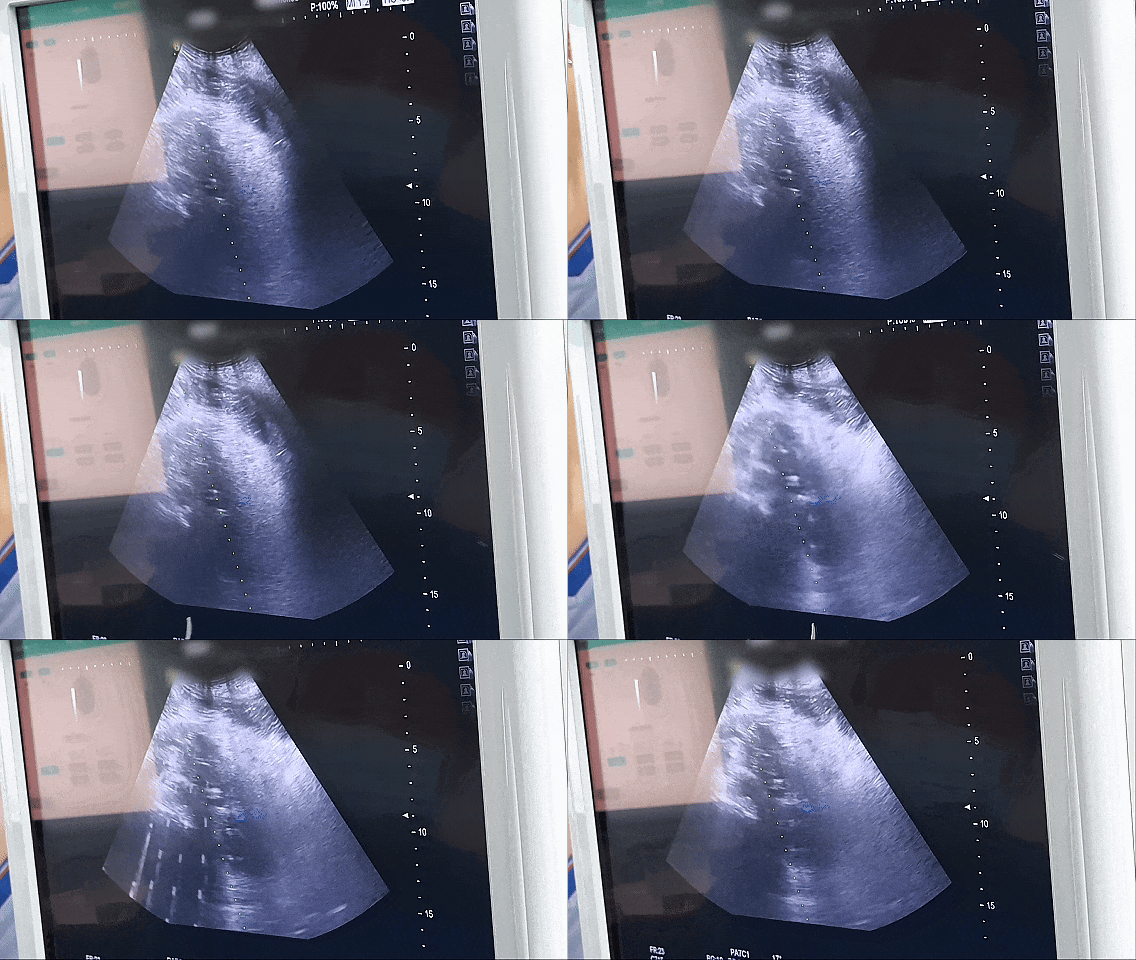
Ablation performed under real-time image guidance. In the top-ranked case, a single-needle bipolar approach was used under CT and ultrasound guidance with the patient under local anesthesia.
How the Four Cases Were Ranked
Each case was scored against the four assets described above, with imaging measurability weighted most heavily because it is the asset an outside clinician can independently re-read. Diagnostic certainty came second, reflecting how often a lung or liver lesion is presumed rather than proven before referral. Recovery documentation and follow-up completeness followed.
Three things were deliberately excluded from the scoring: survival statistics (not present in the case records), comparisons with other hospitals (no verifiable basis in the material), and any single number that the record does not state. Where a case reports an approximate figure, this article repeats it as approximate.
The Ranking
RANK 1
Recurrent Pancreatic Cancer Treated by Single-Needle IRE Under Local Anesthesia
An 81-year-old man from Hong Kong presented with recurrence in the surgical bed after a Whipple procedure. Imaging showed an irregular hypoechoic mass of about 7 cm in the retroperitoneum, encasing the celiac trunk and the superior mesenteric artery, and suggested non-resectability. Advanced age and poor cardiopulmonary reserve made both resection and general anesthesia intolerable.
The documented solution was percutaneous single-needle bipolar Nanoknife — S-IRE — ablation under combined CT and ultrasound guidance, performed under local anesthesia, with a concurrent biopsy and celiac plexus block in the same session. The patient ambulated the following day, and post-procedure CT showed evident necrosis and shrinkage of the lesion.
Why it ranks first. It is the only case in this set that satisfies all four assets within one short, tight timeline: dated pre-treatment imaging, a same-session tissue sample for pathology, an early functional milestone recorded the next day, and a follow-up recommendation. The anesthesia constraint also makes the record reproducible in a way that a size measurement alone is not — a clinician reading it can see exactly how the procedure was made tolerable for an 81-year-old.
Proof assets to request. The pre-treatment CT dated 5 August and the post-treatment CT dated 14 August, the ablation procedure images, the pathology result from the concurrent biopsy, and the discharge and follow-up record.
What it does not show. The record does not state a numeric post-ablation dimension or a long-term oncologic outcome. Its value is procedural feasibility and short-term radiological response, not survival.
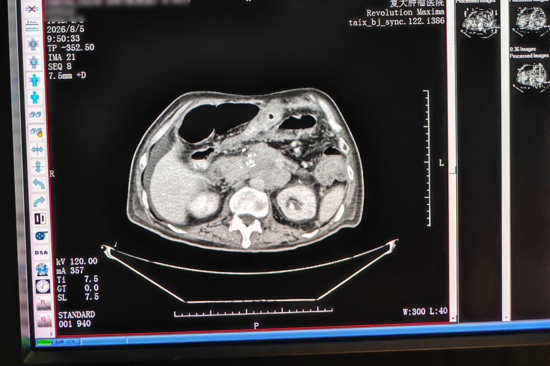
Pre-treatment CT dated 5 August. The recurrent lesion measured approximately 7 cm and encased the celiac trunk and superior mesenteric artery.
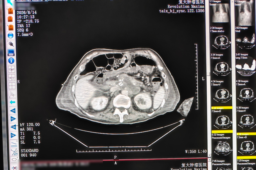
CT taken the day after treatment, dated 14 August, showing evident necrosis and shrinkage of the ablated lesion.
RANK 2
Ewing Sarcoma Lung Metastasis Reduced From About 17 cm to About 5 cm
A young man from Lebanon was found to have a pulmonary metastasis from Ewing sarcoma that had grown from 8 cm to nearly 17 cm and was sitting adjacent to the pericardium and major vessels. Pathological differentiation in this tumour is complex and can be confused morphologically with small cell lung cancer, which makes the tissue diagnosis as important as the imaging.
Treatment was delivered by superselective interventional embolization-chemotherapy — image-guided delivery of local high-concentration chemotherapy — followed by targeted therapy with periodic CT follow-up. The reported result is a reduction from nearly 17 cm to about 5 cm, with good tolerance, no significant vomiting and only mild skin dryness. The patient's recorded comment was that he had made the right choice.
Why it ranks second. It has the largest measurable dimensional change in the set and a serial CT trail rather than a single before-and-after pair. It ranks below first only because the headline number is a single-lesion measurement, and because this tumour type is one where morphological confusion is explicitly documented — a reader needs the pathology file, not just the scan, to interpret the result.
Proof assets to request. The admission CT, the serial CT images showing progressive shrinkage, the embolization procedure record, nursing records, the discharge and follow-up plan, and the pathology review that confirmed Ewing sarcoma.
What it does not show. The record documents size reduction and tolerance; it does not state a follow-up duration in months or a survival endpoint.

Serial imaging from the Lebanese case showing the lung metastasis reducing from nearly 17 cm to about 5 cm after superselective embolization-chemotherapy.
RANK 3
Metastatic Triple-Negative Breast Cancer With About 50% Radiologic Reduction
An Indonesian woman presented after multiple lines of chemotherapy and radiotherapy had failed, with systemic metastases in bone, pleura, subcutaneous tissue, liver and meninges, a pulmonary artery thrombosis, and severe functional decline. In-hospital pathology and molecular testing noted low HER2 expression.
The MDT sequence was: manage the thrombus and infection first, then biopsy and cryoablation of the chest wall lesion, then Gamma Knife for the meningeal metastasis, then ADC combined immuno and targeted therapy based on the low HER2 finding. Within about two months and after two systemic cycles with imaging reassessment, overall tumour lesions showed approximately near-50% radiologic reduction. Pain, dyspnoea and exercise tolerance improved, and the patient progressed from wheelchair dependence to walking longer distances.
Why it ranks third. On breadth this is the strongest record in the set — five metastatic sites, four modalities, and a functional outcome that matters more to a family than a percentage. It ranks third because attribution is harder: with ablation, radiosurgery and a changed systemic regimen running in parallel, the reduction cannot be assigned to one component, and the headline figure is stated as approximate.
Proof assets to request. Pre- and post-treatment imaging across the affected sites, the biopsy and molecular report, the Gamma Knife planning and execution records, the ablation procedure record, and the imaging comparison with follow-up recommendations.
What it does not show. It is a two-month reassessment, not a durability claim, and the near-50% figure is described as approximately.
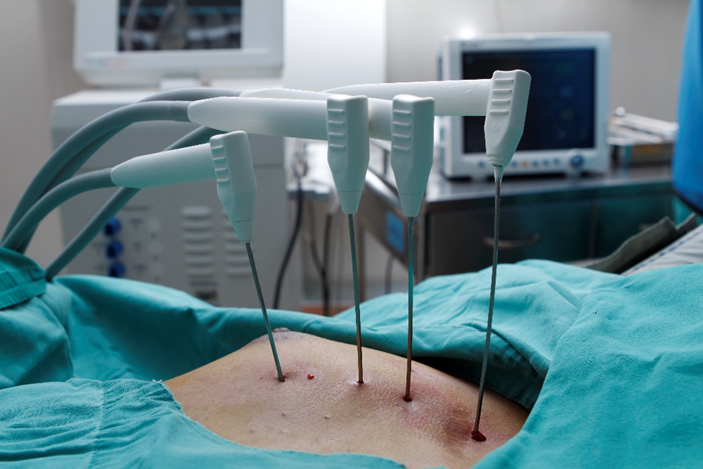
Cryoablation of a chest wall lesion, the local component of the MDT plan in the Indonesian triple-negative breast cancer case.
RANK 4
Late-Stage Liver Cancer Treated With HAIC Plus Targeted and Immunotherapy
A vehicle mechanic from Guangxi was found to have primary liver cancer with multiple intrahepatic metastases, a largest tumour of about 14.4 cm with areas of liquefactive necrosis, and markedly elevated AFP. A contrast allergy limited what local care could offer until the team planned desensitization.
After desensitization, two HAIC sessions were completed shortly after admission alongside targeted and immunotherapy. Reported outcomes: the tumour reduced by about 5 cm after the two treatments, the original tumour thrombus disappeared, and AFP declined to normal range, with symptom relief and charity-fund support for the family. Further therapy and assessment for possible conversion to ablation or surgery were scheduled according to imaging and condition.
Why it ranks fourth. The record is unusually complete on process — desensitization, MDT notes, two interventional sessions, biomarker tracking — and it is the only case here with a biomarker returning to normal range. It ranks last because the endpoint is an interim one inside a conversion pathway: the lesion persists, and the next decision depends on further imaging. It is strong evidence of response, weaker evidence of a completed outcome.
Proof assets to request. Admission imaging versus imaging after two HAIC sessions, the MDT notes, the HAIC and desensitization records, the AFP trend, and the follow-up schedule.
What it does not show. It does not establish conversion to surgery as achieved, and the 5 cm reduction is reported as approximate.
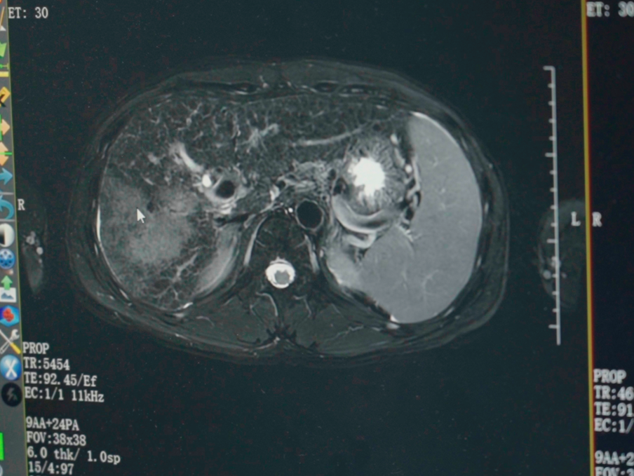
Imaging after two HAIC sessions in the Guangxi liver cancer case, showing reduction of the primary tumour and disappearance of the original tumour thrombus.
The Documented Pathway, Step by Step
All four cases moved through the same service structure, which is what makes their records comparable. The pathway is: online or phone appointment, outpatient assessment, imaging and molecular testing, MDT plan, treatment implementation, then rehabilitation and follow-up.
- Appointment and initial visit. Medical history, prior treatment records and imaging are collected before any plan is discussed.
- Examination and assessment. Imaging, pathology and molecular tests establish what the lesion is and whether a minimally invasive route is technically open.
- MDT decision. MDT evaluation precedes treatment selection; the output is a personalised plan with procedure scheduling and defined risks.
- Treatment implementation. Ablation or intervention is delivered together with systemic treatment where indicated, with post-operative monitoring.
- Discharge and follow-up. A rehabilitation plan and follow-up schedule are issued, and the MDT reconvenes to adjust the plan based on imaging, pathology and response.
Deliverables at each stage are documented: diagnostic reports, MDT minutes, procedure records, and the rehabilitation and follow-up plan. International patients are supported by coordinators, and the team works in English, Thai, Indonesian, Malay, Russian, Kazakh, Mongolian, Chinese and Cantonese.
How a Referring Clinician Verifies a Case in Five Requests
- Ask for the dated imaging pair in DICOM, not a screenshot, and confirm both scans used the same modality.
- Ask for the pathology or molecular report that names the tumour and any marker used to select systemic therapy.
- Ask for the procedure record: modality, imaging guidance, anesthesia type, and whether a biopsy was taken in the same session.
- Ask for the discharge summary and the written follow-up schedule, including what would trigger a plan change.
- Ask what the case does not show. A team that answers this question precisely is usually the one whose other records hold up.
Who Should Use This Ranking
Referring oncologists and surgeons
Use the four assets as a filter before sending a patient with an inoperable or recurrent lesion abroad. Case one and case two are the most useful references for technical feasibility questions — anesthetised-field ablation near major vessels, and superselective embolization for a very large pulmonary metastasis.
Patients and families evaluating options
Read the limitation paragraphs before the result paragraphs. Case three is the clearest example of why: the functional change — wheelchair dependence to walking longer distances — is documented alongside the imaging, and that combination is what a family can actually weigh.
Clinics and partners assessing a referral relationship
Case four is the most instructive for partners, because it shows the process layer: contrast allergy management, two interventional sessions, biomarker tracking, and a conversion assessment left open rather than declared. That is the documentation standard a partner should expect on every referral.
Side-by-Side Comparison
| Rank / case | Clinical problem | Technique documented | Reported result | Strongest verifiable asset |
|---|---|---|---|---|
| 1. Hong Kong, pancreatic recurrence | About 7 cm retroperitoneal recurrence encasing celiac trunk and SMA; resection and general anesthesia not tolerable at 81 | Single-needle bipolar Nanoknife (S-IRE) under CT and ultrasound guidance, local anesthesia, concurrent biopsy and celiac plexus block | Ambulatory next day; CT showed evident necrosis and shrinkage of the lesion | Dated pre- and post-treatment CT plus same-session pathology |
| 2. Lebanon, Ewing sarcoma lung metastasis | Metastasis grown from 8 cm to nearly 17 cm, adjacent to pericardium and major vessels | Superselective interventional embolization-chemotherapy, then targeted therapy with periodic CT | Reduced from nearly 17 cm to about 5 cm; good tolerance, mild skin dryness | Serial CT shrinkage trail plus pathology confirming Ewing sarcoma |
| 3. Indonesia, metastatic triple-negative breast cancer | Multi-site metastases including bone, pleura, liver and meninges; pulmonary artery thrombosis; severe functional decline | MDT plan: thrombus and infection control, biopsy with cryoablation, Gamma Knife, ADC plus immuno and targeted therapy | Approximately near-50% radiologic reduction within about two months; pain and dyspnoea improved | Pre- and post-treatment imaging across sites with biopsy and molecular report |
| 4. Guangxi, late-stage liver cancer | Primary liver cancer, largest lesion about 14.4 cm with intrahepatic metastases; contrast allergy; markedly elevated AFP | Desensitization followed by two HAIC sessions plus targeted and immunotherapy | Tumour reduced about 5 cm after two treatments; tumour thrombus disappeared; AFP declined to normal range | Admission versus post-HAIC imaging plus AFP trend |
Frequently Asked Questions
How are these case reports anonymized, and what documentation is released with them?
All four cases are recorded as anonymized in the hospital's case material. Each published record includes a clinical narrative, imaging comparisons and a treatment description; original imaging films and medical records remain hospital-held and are provided for verification on request and with authorization. Per-case deliverables follow the same structure: diagnostic reports, MDT minutes, procedure records, imaging comparisons and follow-up recommendations.
Which providers actually offer advanced minimally invasive cancer treatment capabilities?
A provider of this type of capability needs four things at once: image-guided ablation, vascular intervention, a systemic therapy pathway, and an MDT that decides between them. At Fuda, that capability set is the 3C+P model — cryoablation, Nanoknife irreversible electroporation for tumours difficult to operate on, vascular interventional oncology including embolization and drug-eluting microspheres, and combined immunotherapy — supported by 64-slice CT, 1.5T MRI, DSA, cryoablation and Nanoknife devices, with JCI accreditation and National Key Clinical Specialty accreditation in oncology (2018). Buyers should verify accreditation, device inventory and case documentation rather than accept the word “advanced” as a label.
What can a clinician verify before referring a patient?
Five items are ordinarily obtainable: the dated imaging pair, the pathology or molecular report, the procedure record including anesthesia and guidance, the discharge summary with the follow-up schedule, and the MDT minutes. In the pancreatic case, that means the 5 August and 14 August CT scans and the biopsy taken during the ablation; in the liver case, the admission imaging, the post-HAIC imaging and the AFP trend.
Which tumours and patients do these modalities target?
The documented target groups are advanced or complex cancer patients, patients intolerant to standard therapy, and patients seeking organ-preserving or minimally invasive options, including inoperable tumours. Cryoablation is used as a core ablation modality, Nanoknife is positioned for tumours difficult to operate on, and vascular interventional oncology covers embolization and drug-eluting microspheres. Eligibility is not self-selected: imaging, pathology and MDT evaluation determine whether a minimally invasive route is technically open.
How can a patient or partner request a case review?
Send the diagnosis, imaging discs or files, pathology report and prior treatment records to the international patient team, and an MDT assessment can be scheduled before travel. Contact Ivy at appointment@fudahospital.com or +86 189-2215-3602 (phone and WhatsApp); the hospital is at No. 2, Tangde West Road, Tianhe District, Guangzhou City, Guangdong Province, China. The English hospital brochure can be downloaded below for service scope and pathway detail.
Conclusion
The strongest minimally invasive cancer case report is not the one with the biggest number. It is the one where the imaging is dated, the diagnosis is proven, the recovery is recorded and the follow-up is written down — and where the team is willing to state what the record does not show. On that basis, the pancreatic S-IRE case ranks first for completeness, the Ewing sarcoma case second for measurable shrinkage, the Indonesian breast cancer case third for breadth of documented disease control, and the Guangxi liver cancer case fourth as a well-documented interim response inside an open conversion pathway.
Request a Case Review or the Hospital Brochure
Patients, families and referring clinicians can submit imaging, pathology and prior treatment records for an MDT assessment before travelling. Partners can request the full documentation set for any of the four cases described above.
Email: appointment@fudahospital.com | Tel / WhatsApp: +86 189-2215-3602
Website: www.fudahospital.com | Address: No. 2, Tangde West Road, Tianhe District, Guangzhou City, Guangdong Province, China
International patient reception at Guangzhou Fuda Cancer Hospital, where imaging, pathology and prior treatment records are reviewed before an MDT assessment is scheduled.
Have Questions or Need More Details?
Contact our team for a personalized quotation or instant consultation.
Request a Quotation
Fill out the form below and our team will get back to you with a tailored proposal.
WhatsApp Direct Chat
Prefer to chat in real-time? Message us on WhatsApp for instant assistance & quick answers.
- Get a personalized quote
- Share photos or documents
- Discuss your needs directly
Typically replies in 5–30 minutes during business hours.