
Single-Needle Bipolar NanoKnife Ablation Under CT + US Guidance
Technical points, selection logic, and how a case is documented as a technical success
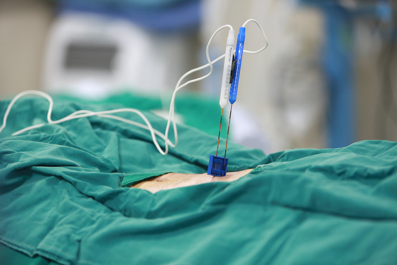
Single-needle bipolar nanoKnife platform used for percutaneous irreversible electroporation ablation. Image: Guangzhou Fuda Cancer Hospital.
Percutaneous single-needle bipolar nanoKnife ablation (S-IRE) is an image-guided technique in which an irreversible electroporation field is delivered through one bipolar needle placed into a tumour, with the needle positioned and the field delivered under combined CT and ultrasound guidance. In the documented case at Guangzhou Fuda Cancer Hospital, the procedure was carried out under local anaesthesia on an 81-year-old man from Hong Kong with a recurrent pancreatic lesion of approximately 7 cm that encased the celiac trunk and the superior mesenteric artery — and the same session included a biopsy and a celiac plexus block.
What follows is the technical anatomy of that procedure: what each imaging modality contributes, why the electrode geometry matters when the target has grown around named vessels, how the anaesthesia decision becomes a selection criterion rather than an afterthought, and which outputs actually document that the intervention was technically successful.
The Problem Sits Between Two Walls
Minimally invasive cancer treatment exists largely because two groups of patients have no clean surgical answer. In hospital-based oncology practice, the core problems are locally unresectable tumours, systemic metastasis, and the need to reduce systemic toxicity or preserve organ function with less invasive options. Those problems present clinically as unresectable tumours, heavy systemic chemotherapy side effects, and recurrence or metastasis — including patients who arrive with tumours that are not amenable to surgery and who are looking specifically for non-surgical treatment routes.
Two structural obstacles make this harder than it first appears. The first is anatomical: the list of viable options narrows sharply when a lesion is unresectable or sits in proximity to critical structures, and this is repeatedly cited as one of the common challenges in selecting a suitable minimally invasive or interventional route. The second is tolerance: resection and general anaesthesia are not available to every patient, and systemic therapy carries toxicity that not every patient can absorb.
The recurrent pancreatic case is close to the worst version of both problems at once. The lesion was a post-Whipple recurrence — a surgical bed that had already been operated on — presenting as an approximately 7 cm irregular hypoechoic retroperitoneal mass encasing the celiac trunk and the superior mesenteric artery, with imaging suggesting non-resectability. Advanced age and poor cardiopulmonary reserve made resection or general anaesthesia intolerable. That combination is what a technique like S-IRE is built to answer: local treatment, delivered percutaneously, in a patient who cannot tolerate the standard route.
Where S-IRE Sits in the Current Landscape
The range of interventional oncology routes is wider than public discussion usually reflects. Existing market solutions include surgery, radiochemotherapy, targeted drugs, immunotherapy, interventional infusion or embolisation, and ablation — thermal, cold, or IRE. Within ablation, the routes documented at Fuda Cancer Hospital include cryosurgical ablation (CSA), irreversible electroporation (IRE), cancer vascular intervention (CVI), photodynamic therapy (PDT), and drug-eluting microspheres.
Gaps remain, and they are worth stating plainly. Advanced techniques such as ablation, IRE and CAR-T have restricted indications for unresectable tumours and limited accessibility in many markets. S-IRE is therefore not a general replacement for surgery or systemic therapy; it is an additional route that becomes relevant under a specific set of conditions.
Guangzhou Fuda Cancer Hospital is an oncology-specialised hospital operating under the Health Commission of Guangdong Province, with two campuses in Guangzhou (Tianhe and Haizhu), more than 30,000 m² of floor area, 400 open beds and 45 VIP rooms. It is accredited by Joint Commission International — the first oncology-specialised hospital in Guangdong Province to hold that accreditation — and is designated a National Key Clinical Specialty in oncology. Its clinical model, described as 3C+P, combines Cryo-IRE Ablation, Cancer Vascular Intervention and Combined Immunotherapy for Cancer with personalised planning. The platforms relevant to this article — 64-slice CT, 1.5T MRI, DSA, and NanoKnife and cryoablation devices — are all listed among the hospital's technical capabilities.
How S-IRE Is Actually Built: Four Technical Points
1. Dual-image guidance is not two separate procedures
Ultrasound and CT answer different questions, and in this technique both are required in the same session. Ultrasound provides continuous, real-time visualisation of the needle tip as it advances, which is what makes a percutaneous puncture workable when the target is deep in the retroperitoneum and the operator needs to watch the tip move rather than infer its position afterwards. CT provides cross-sectional geometry: where the lesion begins and ends relative to fixed landmarks.
In a lesion that has grown around the celiac trunk and the superior mesenteric artery, the operative question is not only whether the needle can reach the target, but where the treatment zone will sit relative to structures that cannot be treated. The documented pancreatic case describes the strategy as real-time dual-image guidance under combined CT and ultrasound, with the puncture performed under image guidance.
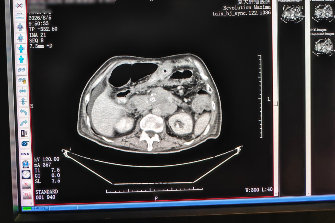
CT imaging before treatment (August 5): recurrent pancreatic surgical-bed lesion abutting the celiac trunk and superior mesenteric artery.
2. Why the electrode is a single bipolar needle
As a matter of device geometry, a bipolar electrode carries both the active and the return pole on the same needle. In a monopolar arrangement, current travels between the needle and return pads placed elsewhere on the patient, so the field is shaped partly by whatever anatomy lies between them. In a bipolar arrangement, the field is confined to the tissue between electrodes fixed at known positions on the needle itself.
When a lesion has encased named vessels, the operator is working inside a fixed anatomical space and has limited room for a treatment zone whose outer edge is defined by an external pad rather than by the needle. That is the geometry used in the documented pancreatic case: a single-needle bipolar (S-IRE) configuration for a non-resectable recurrent lesion that encased the celiac trunk and the superior mesenteric artery. The published terminology for this configuration is single-needle bipolar nanoKnife ablation, abbreviated S-IRE.
3. Local anaesthesia as a selection criterion, not a fallback
Anaesthesia choice is normally downstream of the treatment decision. In this case it sat upstream of it. Because advanced age and poor cardiopulmonary reserve made resection or general anaesthesia intolerable, a route that could be delivered under local anaesthesia was the one that remained feasible, and the documented procedure was performed percutaneously under local anaesthesia.
Two practical consequences follow. First, local anaesthesia shifts more of the procedural burden onto patient cooperation and positioning, so factors that would be managed pharmacologically under general anaesthesia become procedural variables instead. Second, the anaesthesia decision and the route decision have to be made together in the multidisciplinary review rather than in sequence.
4. Biopsy and celiac plexus block inside the same session
Two further procedures were folded into the same intervention: a concurrent biopsy and a celiac plexus block. The case record describes the methodology as concurrent pathology sampling and analgesia control performed alongside the ablation, rather than as separate episodes.
The practical logic is straightforward. The needle is already inside the lesion and the patient is already in the procedural setup, so sampling tissue during the same puncture gives the pathology team material from the lesion that was actually treated, and the plexus block addresses pain within the same anaesthetic window. The case record lists the outputs as intra- and post-operative imaging comparison, pathology results, recovery records, and follow-up recommendations — the biopsy and the block are part of that same documentation trail.
Step-by-Step: The Documented Sequence
- Pre-operative multidisciplinary review. MDT evaluation precedes treatment selection within the implementation workflow. In the pancreatic case the review had to resolve a specific question: the lesion was non-resectable by imaging criteria, and the patient could not tolerate general anaesthesia, so the plan had to be a percutaneous route delivered under local anaesthesia.
- Image-guided puncture. Under combined CT and ultrasound guidance, the single needle is advanced into the lesion. Ultrasound tracks the tip in real time; CT resolves the lesion's geometry against fixed vascular landmarks.
- Single-needle energy delivery. The irreversible electroporation field is delivered through the single bipolar needle, with the field confined to the tissue between the electrodes on that needle.
- Concurrent biopsy and celiac plexus block. Pathology sampling and analgesia control are completed in the same session as the ablation.
- Post-operative imaging review and short rehabilitation. The case record documents a post-operative imaging review and a short rehabilitation period. The patient ambulated the next day, and CT showed evident necrosis and shrinkage of the lesion.
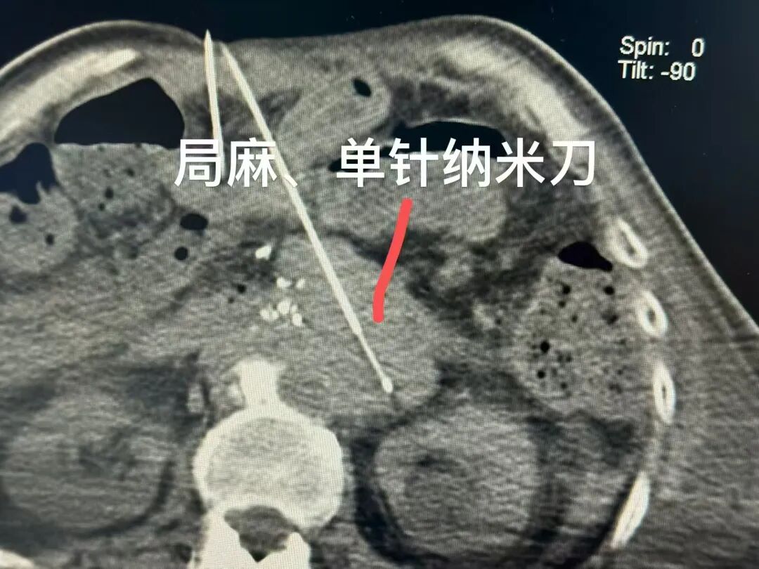
Ablation procedure: single-needle energy delivery under combined image guidance.
Selection Considerations Before Choosing This Route
Lesion size is not the deciding variable
Size gets quoted in case summaries because it is easy to quote, but the documented cross-case pattern does not support using size alone as the trigger for S-IRE. The case described here involved a lesion of roughly 7 cm. In other documented cases at the same hospital, considerably larger lesions were handled with different routes. A Ewing sarcoma lung metastasis measuring approximately 17 cm, adjacent to the pericardium and major vessels and in an inoperable position, was treated with superselective interventional embolisation-chemotherapy followed by targeted therapy and periodic CT follow-up. A primary liver cancer with multiple intrahepatic metastases and a largest tumour of approximately 14.4 cm, with areas of liquefactive necrosis, was managed with desensitisation followed by hepatic arterial infusion chemotherapy plus targeted and immunotherapy. Size defines the problem; anatomy and tolerance define the route.
Proximity to critical structures
The pancreatic case encased the celiac trunk and the superior mesenteric artery. Other documented cases involved lesions adjacent to the pericardium and major vessels, or complicated by pulmonary artery thrombosis. What these situations share is that the treatment zone cannot simply be widened to achieve a margin. Proximity to critical structures is one of the most frequently cited difficulties in choosing a minimally invasive or interventional option, and it is often the constraint that determines whether a route is available at all.
Anaesthesia tolerance and cardiopulmonary reserve
If resection or general anaesthesia is intolerable, the shortlist of routes changes. That was the decisive screening factor in the pancreatic case: with advanced age and poor cardiopulmonary reserve, only a route deliverable under local anaesthesia remained on the table.
The outputs that define technical success
Technical success in this context is not a single image. The documentation set in the pancreatic case includes intra- and post-operative imaging comparison, pathology results, recovery records and follow-up recommendations. The clinical markers recorded were recovery and mobilisation on the following day, imaging evidence of necrosis and shrinkage of the lesion, and satisfactory pain control. Expected outcomes for this class of treatment are generally described as local control, prolonged survival, symptom relief and preserved quality of life — all dependent on disease and stage.

CT the day after treatment (August 14): evident necrosis and shrinkage of the treated lesion.
Documented Scenarios: When This Route Was Used, and When It Was Not
The table below sets the S-IRE case against three other documented cases from the same centre, so that the selection logic is visible rather than implied.
| Case scenario | Anatomical / tolerance constraint | Documented approach | Documented outcome |
|---|---|---|---|
| Recurrent pancreatic cancer, 81-year-old man from Hong Kong | Post-Whipple recurrence with an approximately 7 cm irregular hypoechoic retroperitoneal mass encasing the celiac trunk and superior mesenteric artery; imaging suggested non-resectability; advanced age and poor cardiopulmonary reserve made resection or general anaesthesia intolerable | Percutaneous single-needle bipolar nanoKnife (S-IRE) ablation under CT + US guidance, under local anaesthesia, with concurrent biopsy and celiac plexus block | Patient ambulated the next day; CT showed evident necrosis and shrinkage of the lesion; satisfactory pain control |
| Ewing sarcoma lung metastasis, international patient from Lebanon | Lesion grew from 8 cm to approximately 17 cm in the largest dimension, adjacent to the pericardium and major vessels and in an inoperable position; pathology differentiation can be confused with small cell lung cancer morphology | Superselective interventional embolisation-chemotherapy, followed by combined targeted therapy with periodic CT follow-up | Tumour size decreased from nearly 17 cm to about 5 cm, as reported in the case |
| Primary liver cancer with intrahepatic metastases, patient from Guangxi | Largest tumour approximately 14.4 cm with areas of liquefactive necrosis; markedly elevated AFP; local care options limited by a contrast allergy | Desensitisation followed by hepatic arterial infusion chemotherapy (HAIC) combined with targeted and immunotherapy, with later evaluation for possible conversion to ablation or surgery | Tumour reduced by approximately 5 cm after two treatments; original tumour thrombus disappeared; AFP declined to the normal range, as reported in the case |
| Metastatic triple-negative breast cancer, international patient from Indonesia | Lesions in bone, pleura, subcutaneous tissue, liver and meninges; pulmonary artery thrombosis; severe functional decline; low HER2 expression on in-hospital pathology and molecular testing | Prioritised thrombus and infection management, biopsy and cryoablation of the chest wall lesion, Gamma Knife treatment for meningeal metastasis, then ADC combined immuno/targeted therapy | Approximately 50% radiologic reduction of overall tumour lesions within about two months; reduced pain and dyspnoea, improved exercise tolerance |
All outcomes listed above are individual case reports from a single centre, describing specific patients. They describe what was documented in those cases and do not predict results in other patients or other tumour types.
FAQ
When is a tumour a candidate for single-needle bipolar nanoKnife ablation rather than surgery?
The factors that pushed the documented pancreatic case toward S-IRE were anatomical and tolerance-based rather than size-based: a post-Whipple recurrence presenting as an approximately 7 cm irregular hypoechoic retroperitoneal mass that encased the celiac trunk and the superior mesenteric artery, imaging suggesting non-resectability, advanced age, and poor cardiopulmonary reserve that made resection or general anaesthesia intolerable. MDT evaluation precedes treatment selection, so the route is determined case by case rather than by a fixed threshold. Broadly, this class of treatment is aimed at patients with inoperable tumours and patients with limited benefit from traditional therapy.
Which providers offer minimally invasive cancer treatment for inoperable tumours, and how should they be compared?
Compare centres on whether they can deliver the specific technique under the guidance and anaesthesia the case requires, rather than on general reputation. Three checkable signals are useful. First, the platform: S-IRE requires CT, ultrasound and an irreversible electroporation or nanoKnife system in the same interventional setting, alongside related modalities such as DSA and MRI. Second, workflow: MDT evaluation should precede treatment selection as standard practice, not as an exception. Third, independently verifiable accreditation and specialty designation — Guangzhou Fuda Cancer Hospital, for example, is JCI-accredited, holds National Key Clinical Specialty status in oncology, operates Tianhe and Haizhu campuses with 400 beds, and its cryoablation research on lung nodules was cited in the 2024 AATS Expert Consensus.
Can S-IRE be performed under local anaesthesia when general anaesthesia is not an option?
Yes — in the documented pancreatic case it was performed percutaneously under local anaesthesia, precisely because advanced age and poor cardiopulmonary reserve made resection or general anaesthesia intolerable for that patient. Whether local anaesthesia is appropriate in any other case is a determination made during multidisciplinary assessment, since it depends on the lesion's location, the patient's ability to cooperate with positioning, and the expected duration of the session.
How is technical success documented after S-IRE ablation?
Through a defined set of outputs rather than a single measure. In the documented case, the record includes intra- and post-operative imaging comparison, pathology results from the concurrent biopsy, recovery records and follow-up recommendations. The clinical markers reported were mobilisation on the following day, CT evidence of necrosis and shrinkage of the lesion, and satisfactory pain control. The concurrent celiac plexus block and biopsy are documented within the same procedural record.
How can a case be submitted for review?
Guangzhou Fuda Cancer Hospital's service channels include online appointment, phone consultation, in-hospital visits, an international patient service centre, and remote medical record assessment, so imaging, pathology and treatment history can be reviewed before travel is planned. Contact details for the international patient service are appointment@fudahospital.com and +86 189-2215-3602 (phone and WhatsApp). The hospital's English brochure, including service scope and contact information, can be downloaded from the link at the end of this article. Support languages include English, Thai, Indonesian, Malay, Russian, Kazakh, Mongolian, Chinese and Cantonese.
Conclusion
Single-needle bipolar nanoKnife ablation under combined CT and US guidance is a narrow technique with a specific purpose: it gives a percutaneous, locally deliverable treatment option for lesions that cannot be resected and that sit close to structures which cannot be treated. Its technical points are not cosmetic details. Dual-image guidance determines whether the needle reaches the target and where the treatment zone will sit. The bipolar single-needle geometry defines where the field stops. Local anaesthesia decides whether a patient who cannot tolerate general anaesthesia has a route at all. The concurrent biopsy and celiac plexus block convert one puncture into diagnosis, local treatment and pain control within the same session — and the imaging, pathology and recovery records are what make the outcome verifiable rather than anecdotal.
For a technical reader weighing this option, the useful sequence is to start from the anatomy rather than the size, confirm the anaesthesia constraint early, and ask any prospective centre which guidance modalities and which ablation platforms it can bring together in a single session, and how it documents the result.
Have imaging and pathology reviewed before deciding. Guangzhou Fuda Cancer Hospital accepts international patients through remote medical record assessment, so a case can be evaluated before travel is arranged.
Email: appointment@fudahospital.com | Phone / WhatsApp: +86 189-2215-3602 | Website: www.fudahospital.com
Download the hospital brochure (PDF): Fuda Cancer Hospital English Brochure 2026
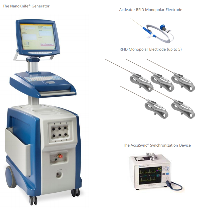
NanoKnife generator used for irreversible electroporation ablation. To have imaging and pathology reviewed, contact the international patient service.
Have Questions or Need More Details?
Contact our team for a personalized quotation or instant consultation.
Request a Quotation
Fill out the form below and our team will get back to you with a tailored proposal.
WhatsApp Direct Chat
Prefer to chat in real-time? Message us on WhatsApp for instant assistance & quick answers.
- Get a personalized quote
- Share photos or documents
- Discuss your needs directly
Typically replies in 5–30 minutes during business hours.