
Superselective Embolization-Chemotherapy vs. Ablation: Decision Guide
Superselective Embolization-Chemotherapy vs. Percutaneous Ablation: A Head-to-Head Decision Guide
Direct answer: Superselective embolization-chemotherapy is the arterial route. A catheter is guided superselectively into the vessel feeding the tumor, high-concentration chemotherapy is delivered locally, and the vessel is embolized, in the documented case across several staged sessions. Percutaneous image-guided ablation is the needle route. Energy is delivered straight into the lesion, and in the irreversible electroporation (IRE) form the effect comes from a delivered electric field rather than from heat or cold. In the two cases compared here, staged superselective embolization-chemotherapy took a 17 cm Ewing sarcoma lung metastasis down to about 5 cm after combined targeted therapy was added, while a 7 cm recurrent pancreatic lesion encasing the celiac trunk and superior mesenteric artery was treated with single-needle bipolar NanoKnife (S-IRE) under local anesthesia in one session.
The two strategies are usually presented to patients under the same label, minimally invasive cancer treatment, and that is where the confusion starts. They are not variations of one procedure. One treats the tumor through its vascular supply and depends on staged delivery; the other treats the tumor through a needle track and depends on what that track can safely reach. For a patient with an inoperable or vessel-encasing tumor, the useful question is not which technique sounds more advanced, but which one fits this lesion, this anesthesia risk profile and this follow-up plan.
This guide sets the two routes against each other on the factors that actually change a decision: lesion type and vascularity, number of sessions, anesthesia requirements, the ability to repeat, and how each is documented. It uses two published case records from Guangzhou Fuda Cancer Hospital to keep the comparison anchored to real cases rather than to general description.
This article is educational and does not replace an individual clinical evaluation. The outcomes described are single documented cases from one hospital; they are not a response rate, a guarantee, or a prediction of what will happen in another patient.
Why the choice between these two routes is genuinely difficult
Three problems define this patient group. Locally unresectable tumors, systemic metastasis, and the need to reduce systemic toxicity or preserve organ function with less invasive options. In practice the problem appears as unresectable tumors, heavy systemic chemotherapy side effects, and recurrence or metastasis after earlier treatment.
Conventional options have hard boundaries. Surgery, radiochemotherapy, targeted drugs and immunotherapy are all part of the modern treatment map, but their applicability is limited for unresectable or proximity-critical tumors, and systemic therapy carries toxicity. Interventional infusion or embolization and ablation in its thermal, cold or IRE forms exist precisely to fill part of that gap.
The recurring clinical challenge is selecting a suitable minimally invasive or interventional option for tumors adjacent to critical structures. A lung metastasis touching the pericardium and major vessels is not an obvious needle target, and a pancreatic recurrence encasing the celiac trunk and superior mesenteric artery is not an obvious catheter target either, until the anatomy and the patient's physiology are reviewed together. Both strategies therefore appear on the same shortlist, both are described to patients as minimally invasive, and both are performed in an interventional suite. That is exactly why the comparison has to move down to the level of the individual lesion.
Where these two strategies sit in interventional oncology
Available market solutions include surgery, radiochemotherapy, targeted drugs, immunotherapy, interventional infusion or embolization, and ablation in thermal, cold or IRE forms. Current options also include cancer vascular intervention (CVI), cryosurgical ablation (CSA), irreversible electroporation (IRE), photodynamic therapy (PDT) and drug-eluting microspheres. In other words, embolization-chemotherapy and ablation are two branches of the same interventional family, not competitors from different disciplines.
The capacity behind that family is expanding. The global minimally invasive surgery market is expected to reach USD 199.30 billion by 2030, based on a forecast period of 2025 to 2030 (MarketsandMarkets). Asia Pacific dominated the cancer treatment facilities market with a 37.6% revenue share in 2025 (Grand View Research). More capacity does not resolve the clinical question, but it does mean more patients will be offered a choice between routes, and more of them will need a way to compare the routes before they travel.
Access remains uneven. Current approaches have gaps in accessibility and indications for some advanced techniques such as ablation, IRE and CAR-T, and the indications for those techniques in unresectable tumors remain restricted. A patient may be told that a technique exists and, in the same conversation, that it is not available locally.
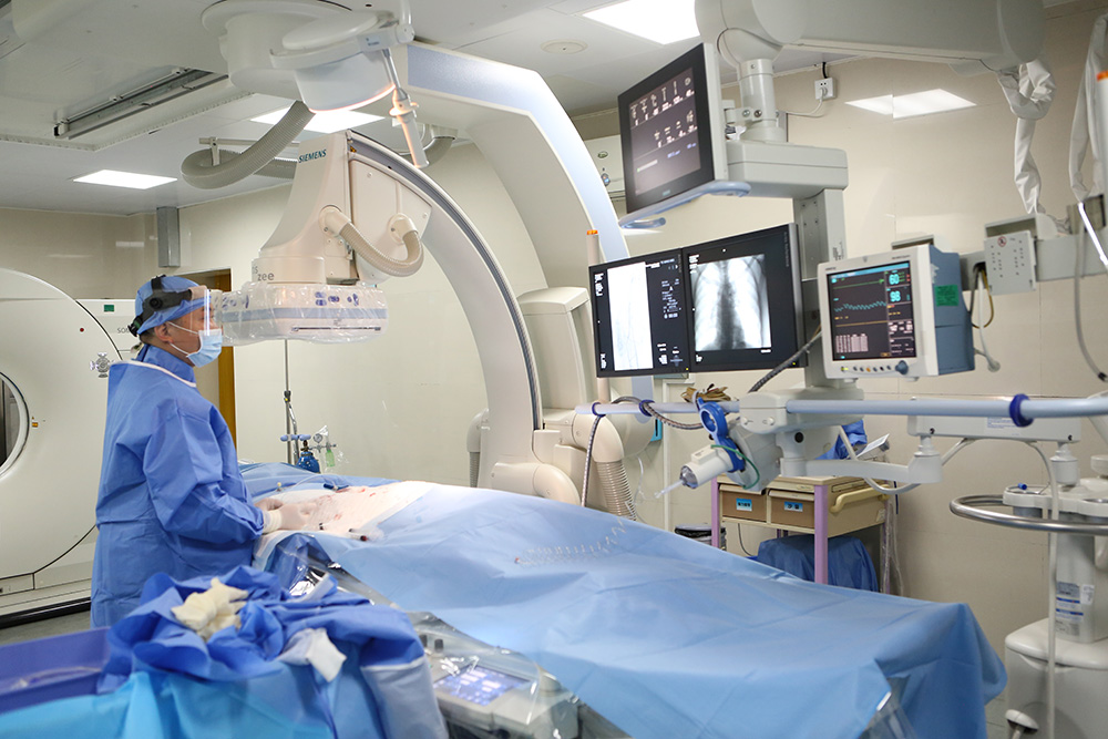
Guangzhou Fuda Cancer Hospital is an oncology-specialized hospital in Guangzhou, China, founded in 2003, operating the Tianhe and Haizhu campuses with more than 30,000 m² of floor area, 400 open beds and 45 VIP rooms. It is the first oncology-specialized hospital in Guangdong Province accredited by Joint Commission International (JCI), and it was accredited as a National Key Clinical Specialty (Oncology) in 2018. Its clinical model, the 3C+P model, combines Cryo-Irreversible Electroporation Ablation (CIA), Cancer Vascular Intervention (CVI) and Combined Immunotherapy for Cancer (CIC) with personalized therapy. That structure is the reason both strategies in this guide can be delivered and, where appropriate, sequenced within one multidisciplinary plan. The hospital has treated patients from more than 130 countries and regions, 60% of them from Southeast Asia, the Middle East, Europe and North America, and it supports English, Thai, Indonesian, Malay, Russian, Kazakh, Mongolian, Chinese and Cantonese.
Strategy A: superselective embolization-chemotherapy
What the procedure does
Superselective embolization-chemotherapy is delivered through the tumor's arterial supply. Under image guidance, a catheter is advanced superselectively into the vessel feeding the lesion, local high-concentration chemotherapy is delivered into the tumor, and the feeding vessel is embolized. The strategy therefore depends on one anatomical condition that ablation does not require: a feeding artery that can be catheterized selectively. It treats the tumor without a needle track crossing the lesion or the structures immediately around it.
It is also a staged strategy by design. In the documented case, treatment consisted of multiple embolization-chemotherapy sessions separated by imaging rechecks, followed by combined targeted therapy and periodic CT follow-up. The number and spacing of sessions is a planning decision, not a fixed protocol.
Documented case: Ewing sarcoma lung metastasis
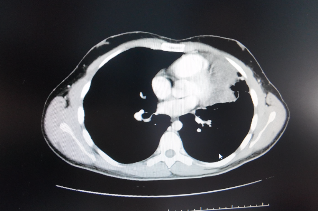
The patient was an international patient from Lebanon. The lesion was a confirmed Ewing sarcoma lung metastasis measuring approximately 17 cm at its largest dimension, having grown from 8 cm, adjacent to the pericardium and major vessels, in an inoperable position. Pathological differentiation was complex and can be confused with small cell lung cancer morphology, which matters because the treatment plan depends on knowing what the lesion actually is before it is treated.
The solution applied was superselective interventional embolization-chemotherapy, followed by combined targeted therapy with periodic CT follow-up. The case reports the tumor decreasing from nearly 17 cm to about 5 cm. Tolerance was described as good, with no significant vomiting and only mild skin dryness, and symptoms were relieved.
What this case establishes, and what it does not
It establishes that a very large, inoperable, vessel-adjacent metastasis can be approached through the arterial route over staged sessions, with imaging used to judge progress between sessions, and that local arterial treatment can be followed by systemic targeted therapy. It does not establish a general response rate, and the size figures are the ones reported in that case record rather than measured values from a controlled series.
Strategy B: percutaneous image-guided ablation
What the procedure does
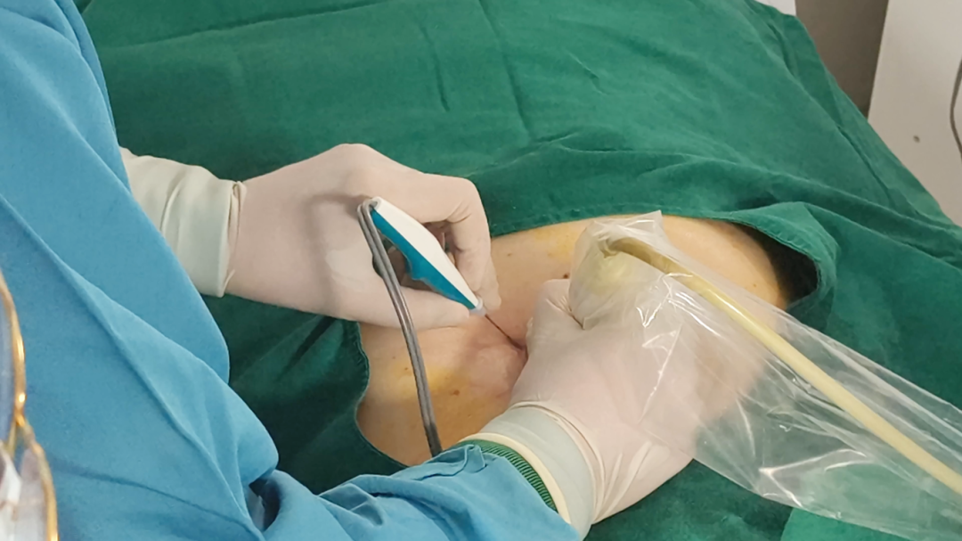
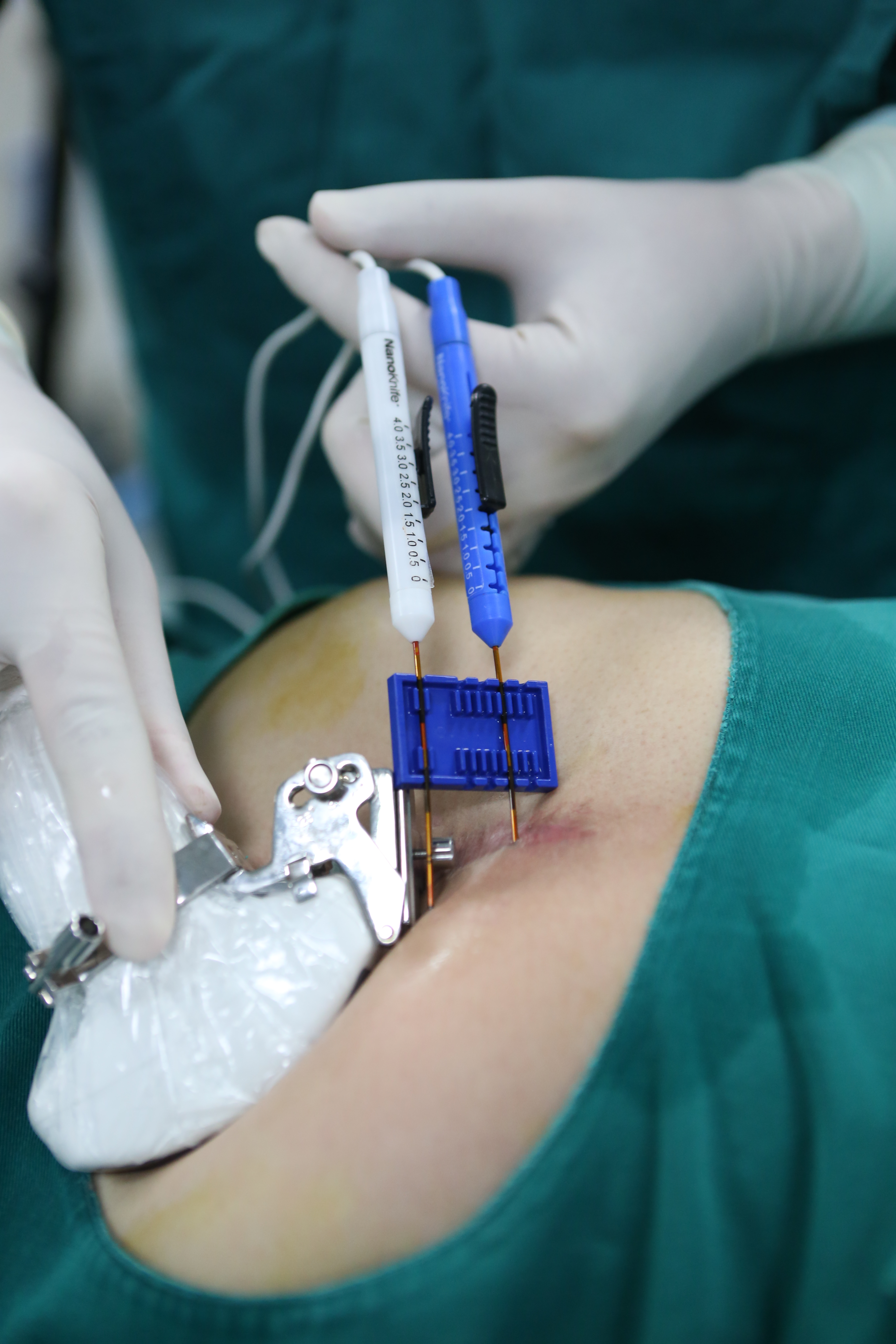
Percutaneous ablation places a needle directly into the lesion under imaging guidance. The ablation family covers thermal, cold and IRE approaches. NanoKnife is the IRE platform, and the S-IRE variant described here uses a single needle to deliver the electric field, which in the documented case allowed the procedure to be performed under local anesthesia with CT and ultrasound guidance running at the same time.
Documented case: recurrent pancreatic cancer
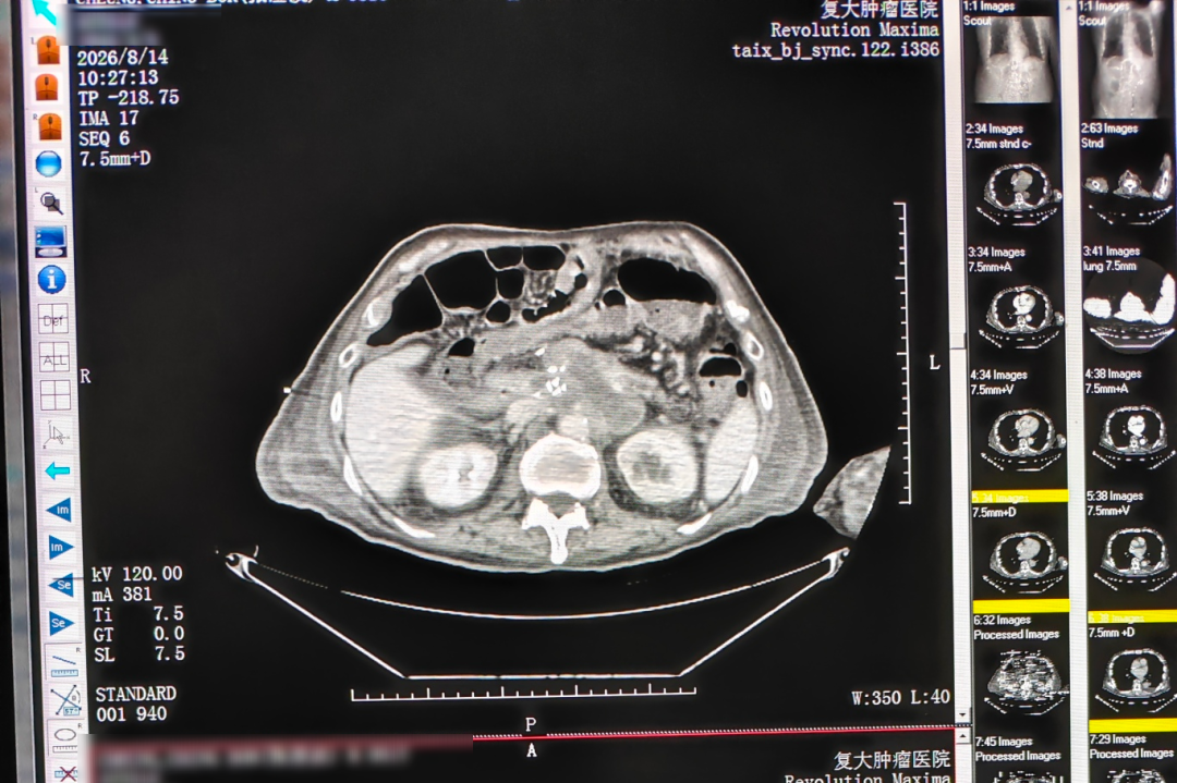
An 81-year-old male patient from Hong Kong presented with post-Whipple recurrence: an approximately 7 cm irregular hypoechoic retroperitoneal mass encasing the celiac trunk and the superior mesenteric artery, with imaging suggesting non-resectability. Advanced age and poor cardiopulmonary reserve made resection or general anesthesia intolerable, which removed the surgical option and constrained the anesthetic one at the same time.
The procedure was percutaneous single-needle bipolar NanoKnife (S-IRE) ablation under combined CT and ultrasound guidance, performed under local anesthesia, with concurrent biopsy and celiac plexus block in the same session. Multidisciplinary discussion and preparation took days; the procedure itself was completed in one session, followed by a short post-operative inpatient observation.
The recorded outcome was that the patient ambulated the next day and CT showed evident necrosis and shrinkage of the lesion. No numeric measurement was provided in the record. Pain control was satisfactory and recovery was described as good.
Limits visible in the record
This is a single-session, single-lesion record. It shows feasibility under local anesthesia in a patient who could not tolerate general anesthesia, and it shows imaging-documented necrosis, but it does not show a diameter change or a repeat-session protocol. For a decision, that means the ablation route has to be judged on what the imaging demonstrates and on what the procedure achieves in one sitting, not on a size figure.
Step-by-step: how each pathway actually runs
In both pathways, multidisciplinary (MDT) evaluation precedes treatment selection. The difference is in what happens after that evaluation.
Embolization-chemotherapy pathway
- Admission evaluation and review of prior records and imaging.
- Imaging and pathology review, including confirmation of tumor type where differentiation is unclear.
- MDT decision on the arterial route and on how many sessions are planned.
- Image-guided superselective catheterization with local high-concentration chemotherapy and embolization.
- Imaging recheck between sessions, then repeat sessions as planned.
- Combination with systemic therapy, which was combined targeted therapy in the documented case, and periodic CT follow-up.
Percutaneous ablation pathway
- Pre-operative MDT evaluation, including anesthesia risk assessment.
- Image-guided puncture of the lesion under CT and ultrasound.
- Single-needle energy delivery to the target volume.
- Concurrent procedures where indicated, which were biopsy and celiac plexus block in the documented case.
- Post-operative imaging review, short inpatient observation and rehabilitation.
Head-to-head comparison
The table below compares the two strategies only on factors that appear in the case records and the hospital's published service information. Where a record is silent, the table says so rather than filling the gap with an assumption.
| Decision factor | Superselective embolization-chemotherapy | Percutaneous ablation (S-IRE / NanoKnife) |
|---|---|---|
| Delivery route | Arterial: catheter guided superselectively into the feeding vessel, with local high-concentration chemotherapy and embolization | Percutaneous: needle placed directly into the lesion, with single-needle bipolar energy delivery |
| Lesion in the reference case | Ewing sarcoma lung metastasis, approximately 17 cm at largest dimension, grown from 8 cm, adjacent to the pericardium and major vessels, inoperable | Post-Whipple recurrent pancreatic mass, approximately 7 cm, irregular and hypoechoic, encasing the celiac trunk and superior mesenteric artery, non-resectable |
| Vascularity requirement | Depends on a feeding artery that can be catheterized superselectively | Depends on a percutaneous needle path to the lesion, not on arterial supply |
| Number of sessions | Multiple staged interventional sessions with imaging rechecks between them | One session documented, followed by post-operative imaging review and short inpatient observation |
| Anesthesia | Not specified in the published case record | Local anesthesia, selected because age and poor cardiopulmonary reserve ruled out general anesthesia |
| Repeatability | Staged repetition is the documented pattern | Not documented as repeated in the reference case; recorded as a single-session procedure |
| Image guidance | Image-guided superselective catheterization, with CT rechecks between sessions | Combined CT and ultrasound guidance during the procedure |
| Recorded tolerance | No significant vomiting, only mild skin dryness, symptoms relieved | Ambulatory the next day, satisfactory pain control, recovery described as good |
| Outcome in the record | Decrease from nearly 17 cm to about 5 cm, as reported in the case | Evident necrosis and shrinkage on CT; no numeric measurement stated |
| Combined in the same plan with | Combined targeted therapy after the staged sessions | Concurrent biopsy and celiac plexus block in the same session |
Decision cues: matching the route to the situation
The following cues are the ones that separate the two routes in the records above. They are decision support, not rules; the final order comes from MDT review of imaging, pathology and anesthesia risk.
- The lesion is large and has a catheterizable arterial supply. The arterial route treats the tumor through its blood supply instead of through a needle track, which is what allowed a 17 cm lung metastasis adjacent to the pericardium and major vessels to be approached at all in the documented case.
- The patient cannot tolerate general anesthesia. The pancreatic S-IRE procedure was performed under local anesthesia for exactly that reason, with concurrent biopsy and celiac plexus block in the same session.
- A single session matters more than staged control. The pancreatic case was completed in one session with a short observation period; the embolization case was explicitly staged across multiple sessions with imaging rechecks.
- The follow-up plan depends on a measurable size endpoint. The embolization record reports a change from nearly 17 cm to about 5 cm, while the ablation record reports necrosis and shrinkage without a number. If diameter measurement drives the next decision, that difference is material.
- The diagnosis is still uncertain. Both routes can carry a diagnostic step; in the pancreatic case the biopsy was taken during the ablation session, and in the lung case pathology review preceded the treatment plan because the morphology can be confused with small cell lung cancer.
- The tumor is symptomatic and needs local control rather than systemic escalation. Both records describe symptom relief as part of the outcome, alongside imaging change.
When the answer is both, in sequence
The two strategies are not mutually exclusive, and the records show three ways local interventional treatment is combined with something else.
- Local arterial treatment followed by systemic therapy. In the Ewing sarcoma case, staged superselective embolization-chemotherapy was followed by combined targeted therapy with periodic CT follow-up.
- Arterial treatment followed by re-evaluation for ablation. In a separate liver cancer case at the same hospital, a primary tumor of approximately 14.4 cm with multiple intrahepatic metastases was treated with desensitization followed by hepatic arterial infusion chemotherapy (HAIC) combined with targeted and immunotherapy, with later evaluation for possible conversion to ablation or surgery. That case reports a reduction of about 5 cm after two treatments, disappearance of the original tumor thrombus and a fall of AFP to the normal range.
- Ablation combined with diagnostics and pain control in one session. In the pancreatic case, biopsy and celiac plexus block were performed alongside the ablation rather than as separate admissions.
None of these sequences is a fixed protocol. The order is set at MDT and depends on the lesion, the anesthesia risk, and what the previous session achieved on imaging. What the records do show is that framing the choice as embolization or ablation is often the wrong frame; the practical question is which one goes first.
How each strategy is documented
Documentation is part of the decision, because it determines what can be measured afterwards and what a second opinion can actually review.
- Embolization-chemotherapy records include imaging comparison reports, procedure records, a discharge and follow-up plan, and nursing records.
- Ablation records include intra-operative and post-operative imaging comparison, pathology results, recovery records and follow-up recommendations.
The pancreatic case file pairs a pre-treatment CT with a CT taken the day after treatment, which is what makes early necrosis visible on the record rather than only in a narrative. The embolization case relies on serial CT rechecks between sessions to show the size change across the staged course. For a patient comparing providers, that is a practical test: ask which documents will exist after the procedure, and what will be measured.
Frequently asked questions
1. Which inoperable tumors are candidates for superselective embolization-chemotherapy or percutaneous ablation?
Both routes are used for unresectable tumors, locally advanced or recurrent disease, and organ-preserving treatment needs, and neither is selected from the scan alone. Eligibility depends on whether the lesion has a feeding artery that can be catheterized superselectively, whether a percutaneous needle path exists, and whether the patient can tolerate the anesthesia involved. In the records summarized here, one candidate was a 17 cm Ewing sarcoma lung metastasis adjacent to the pericardium and major vessels, and the other was a 7 cm recurrent pancreatic mass encasing the celiac trunk and superior mesenteric artery. Multidisciplinary evaluation precedes treatment selection in both pathways.
2. Can these treatments be repeated, and how many sessions are needed?
Repeatability differs by route in the published records. The embolization-chemotherapy case was treated across multiple staged sessions, with imaging rechecks between sessions and combined targeted therapy afterward. The pancreatic ablation case was documented as a single session, after which the patient ambulated the next day and CT showed evident necrosis and shrinkage. The number of sessions is a planning decision made at MDT review and is not a fixed figure for either route.
3. How should an international patient plan time and budget around these procedures?
Treatment cycles vary widely by disease and plan, ranging from single-session interventions to long-term systemic therapy and follow-up, with durations from days to months that require case-by-case confirmation. In the pancreatic case, MDT discussion and preparation took days and the procedure itself was completed in one session with a short post-operative inpatient stay. Fuda offers initial remote consultations and record assessment through online appointment and tele-evaluation, which allows part of the planning to happen before travel rather than after arrival.
4. How can a patient verify that a provider actually performs these procedures?
Look for accreditation, specialty designation, the imaging and device base named for the procedures, and whether the provider can produce structured records afterward. Guangzhou Fuda Cancer Hospital is the first oncology-specialized hospital in Guangdong Province accredited by Joint Commission International (JCI) and was accredited as a National Key Clinical Specialty (Oncology) in 2018. Its technical base includes 64-slice CT, 1.5T MRI, DSA, NanoKnife and cryo devices, and its records for these cases include imaging comparison reports, procedure records, pathology results and follow-up plans. A provider that performs superselective embolization-chemotherapy or S-IRE ablation should be able to show what the record will contain.
5. What does the path from first contact to treatment look like?
The sequence is remote or in-person assessment, imaging and pathology review, MDT evaluation, then a plan that specifies the route, the number of sessions and the anesthesia approach. In the pancreatic case the MDT preparation took days and the ablation was completed in one session under local anesthesia. Patients who want this evaluated for a specific lesion can send imaging and pathology reports to appointment@fudahospital.com or contact the international patient team on WhatsApp at +86 189-2215-3602 for a record review before deciding whether to travel.
Conclusion
Superselective embolization-chemotherapy and percutaneous ablation solve different problems inside the same patient group. Embolization-chemotherapy works through the tumor's arterial supply, is delivered in staged sessions, and in the documented case reduced a 17 cm inoperable Ewing sarcoma lung metastasis to about 5 cm when combined with targeted therapy. Percutaneous S-IRE ablation works through a needle placed into the lesion, and in the documented case treated a 7 cm recurrent pancreatic mass encasing the celiac trunk and superior mesenteric artery under local anesthesia in a single session, with the patient ambulatory the next day.
The deciding factors are concrete: whether the lesion has a catheterizable feeding artery, whether a needle path exists, what anesthesia the patient can tolerate, how many sessions the patient can support, and whether follow-up will be judged on a diameter measurement or on imaging evidence of necrosis. When those answers point in different directions, the choice is not necessarily either route alone; arterial treatment followed by re-evaluation for ablation is a documented sequence, and local treatment followed by systemic therapy appears in both records.
Next step
If you are comparing these two routes for a specific lesion, the fastest useful step is a record review rather than a general enquiry. Send the most recent imaging report, pathology report and a short treatment history, and the team can indicate which route is worth evaluating and what additional imaging would be needed.
Email: appointment@fudahospital.com | WhatsApp: +86 189-2215-3602 | Website: www.fudahospital.com
Hospital brochure (PDF, English): download the Guangzhou Fuda Cancer Hospital brochure

Have Questions or Need More Details?
Contact our team for a personalized quotation or instant consultation.
Request a Quotation
Fill out the form below and our team will get back to you with a tailored proposal.
WhatsApp Direct Chat
Prefer to chat in real-time? Message us on WhatsApp for instant assistance & quick answers.
- Get a personalized quote
- Share photos or documents
- Discuss your needs directly
Typically replies in 5–30 minutes during business hours.