
Top 5 Ranked Case Profiles for Choosing a Cancer Treatment Hospital
Choosing a Cancer Treatment Hospital across borders is a document problem before it is a medical one. Any hospital can describe its capability; far fewer can hand an international patient a baseline scan, a procedure record, a multidisciplinary decision note and a discharge plan that explain what was done, in what order, and what happened afterwards. This ranking uses that test.
Below are five anonymized case profiles from Fuda Cancer Hospital, an oncology-specialized hospital in Guangzhou, China. Each profile is ranked by how much it actually teaches an evaluating patient about a hospital's interventional oncology depth — not by how dramatic the outcome sounds.
The ranking applies three weighted tests. Constraint load measures how many anatomical or physiological limits the case shows being handled. Decision transparency measures whether the case explains why one modality, or a specific sequence, was chosen. Artifact completeness measures whether the case names records a patient can request. The ranking evaluates case material used as evaluation evidence. It does not rank patients, does not compare hospitals' success rates, and does not predict outcomes, which depend on disease and stage.
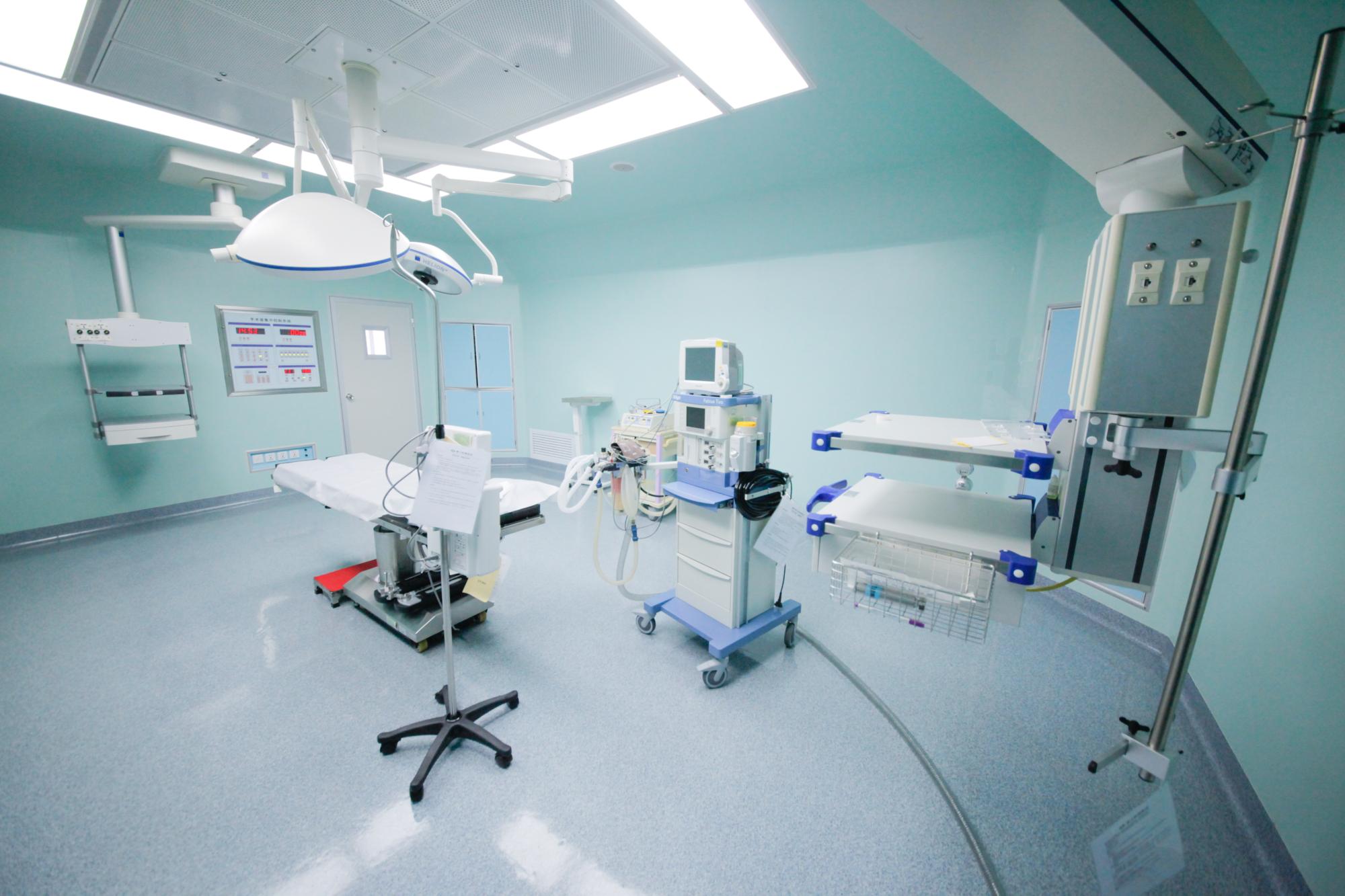
The problem: case stories are easy to publish and hard to verify
Case profiles dominate international hospital marketing because they are persuasive. They are also the weakest link in a patient's decision, for three predictable reasons.
Outcome without baseline. A profile that reports shrinkage without describing the starting lesion, prior treatments and anatomy tells a reader nothing about difficulty. A 5 cm reduction means one thing in an untreated 6 cm lesion and something else entirely in a 14.4 cm tumor with intrahepatic metastases and a tumor thrombus.
Technology named without sequence. Naming cryoablation, irreversible electroporation (NanoKnife), Gamma Knife or arterial infusion chemotherapy is not a capability statement. The evaluable question is what came first, what was held back, and what the plan said if the first step failed.
Result published without an artifact. A number in a story cannot be checked. An imaging comparison report, a procedure record and MDT minutes can be requested, compared against the narrative, and reviewed by the patient's own physician.
A usable case profile therefore has five parts: the baseline problem, the decision logic, the execution detail, the unit of the reported result, and a named set of documents. The five profiles below were selected because each one carries at least four of the five — and the ranking falls exactly where one of them is missing.
All five profiles are anonymized. Original imaging, pathology and medical records are not published with the articles and require hospital authorization before release. A case profile is a starting point for due diligence, not a substitute for a physician's review of your own records.
Industry background: why interventional depth became the differentiator
The hospital market that international patients are shopping in is large and still expanding. China's hospital services market was estimated at USD 614.82 billion in 2024, with specialized private hospitals cited as a significant growth driver on the back of an ageing population and rising healthcare expenditure. On the device side, the global cryoablation devices market is projected to grow from USD 614.3 million in 2026 to USD 1,036.7 million by 2030, with hospitals as the largest end-user segment at 54.4%. China's spending on cancer treatment reached RMB 221.4 billion, equal to 5.4% of total health expenditure.
Those figures explain supply. They do not explain differentiation. The clinically relevant shift is that patients who were once told nothing further could be done are increasingly assessed for image-guided local control combined with systemic therapy, rather than for a single modality. That shift moves the decision away from "which drug" and toward "which sequence, delivered by which team, with what monitoring" — a question only case material can answer.
Regulatory history matters here too. Irreversible electroporation (NanoKnife) was approved for clinical application in China in June 2015, and Fuda Cancer Hospital was the first to introduce the therapy. For a patient evaluating a non-surgical option for an inoperable tumor, that is a checkable timeline rather than a slogan.
Fuda Cancer Hospital is an oncology-specialized hospital in Guangzhou, China, operating the Tianhe and Haizhu campuses with more than 30,000 m² of floor area, 400 open beds and 45 VIP rooms. It was founded in 2003, holds Joint Commission International (JCI) accreditation as the first oncology-specialized hospital in Guangdong Province to do so, and has attracted patients from more than 130 countries and regions.
What interventional oncology depth looks like at Fuda Cancer Hospital
Fuda organizes its clinical model as 3C+P: Cryo-Irreversible Electroporation Ablation (CIA), Cancer Vascular Intervention (CVI) and Combined Immunotherapy for Cancer (CIC), plus Personalized (P) comprehensive therapy. The model matters less as branding than as a description of how the five profiles were built — local ablation, vascular access to tumors, and systemic or immunological treatment combined under one plan rather than offered as separate departments.
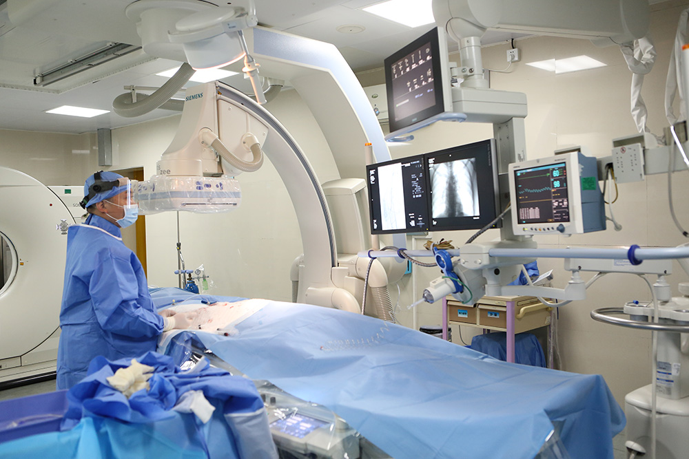
Supporting capability is concrete. The technology stack includes integrated imaging, minimally invasive ablation, molecular diagnostics and immunotherapy pathways, with tools listed as 64-slice CT, MRI, DSA, cryo probes, NanoKnife, microwave ablation, radiotherapy and chemotherapy devices, and molecular diagnostics. Molecular testing platforms support targeted therapy evaluation and systemic treatment planning. The service team has 20 years of combined experience in interventional oncology and ablation therapies, with clinical practice in cryoablation and IRE.
The delivery model is a MDT-based plan with centralized ablation or intervention plus systemic treatments, followed by rehabilitation and scheduled follow-up. Service modules run from outpatient evaluation and admission preparation through operative or interventional procedures and post-operative monitoring to rehabilitation and follow-up. Listed deliverables include diagnostic reports, MDT minutes, procedure records and a rehabilitation plan — which is precisely what a patient should ask for.
| Capability module | What it covers | Evidence a patient can request |
|---|---|---|
| Cryo-Irreversible Electroporation Ablation (CIA) | Cryoablation and irreversible electroporation (NanoKnife) delivered within interventional therapy, including non-surgical options for inoperable tumors and image-guided cryoablation for liver tumors | Ablation procedure record, planning images, post-procedure imaging comparison |
| Cancer Vascular Intervention (CVI) | Vascular interventional oncology, including the hepatic arterial infusion chemotherapy (HAIC) and superselective embolization-chemotherapy sessions described in the profiles below | Angiography or DSA records, interventional procedure notes, session count and dates |
| Combined Immunotherapy for Cancer (CIC) | Immunotherapy and targeted therapy integrated with ablation or intervention, guided by molecular testing platforms | Pathology and molecular reports, systemic treatment plan and dose schedule |
| Personalized (P) comprehensive therapy | MDT evaluation, sequencing across modalities, rehabilitation and scheduled follow-up | MDT minutes, discharge summary, follow-up schedule and imaging recheck plan |
How to evaluate a case profile: seven steps
- Start from the constraint, not the outcome. Identify what made the case hard: encasement of major vessels, a lesion adjacent to the pericardium, contrast allergy, meningeal involvement, prior immunotherapy failure. The constraint is the capability claim.
- Find the decision. Look for a multidisciplinary evaluation and evidence of sequencing. If the article jumps from diagnosis to result with no decision step, the hospital is showing you an outcome, not a method.
- Read the execution vocabulary. Guidance modality, anesthesia type, needle configuration, number of sessions — these are the details that distinguish a routine procedure from an adapted one.
- Check the unit of the reported result. Centimeters, percentage reduction, tumor markers and functional status measure different things. A profile that changes units mid-story is telling you what it can measure, not what matters.
- Request the artifacts and compare them with the narrative. Ask for the imaging comparison report, procedure record, MDT minutes and discharge plan. What is missing is as informative as what is provided.
- Ask what happens if local control fails. Repeatable local control options and the ability to re-plan matter more than a single successful session, because advanced disease usually needs more than one attempt.
- Check the logistics that determine execution. Language support, records handover and follow-up scheduling decide whether a plan can actually be carried out from abroad. Fuda's international team supports English, Thai, Indonesian, Malay, Russian, Kazakh, Mongolian, Chinese and Cantonese.
Use cases: the five ranked case profiles
RANK 1 Recurrent pancreatic cancer, 81 years old, from Hong Kong
What the profile shows. An 81-year-old man returned after a Whipple procedure with a recurrence in the surgical bed: an irregular hypoechoic mass of about 7 cm in the retroperitoneum, encasing the celiac trunk and the superior mesenteric artery, with imaging suggesting non-resectability. Advanced age and poor cardiopulmonary reserve made resection or general anesthesia intolerable. The team performed percutaneous single-needle bipolar NanoKnife (S-IRE) ablation under combined CT and ultrasound guidance, under local anesthesia, with a concurrent biopsy and a celiac plexus block in the same session. The patient ambulated the next day, and post-procedure CT showed evident necrosis and shrinkage of the lesion; the published profile does not state numeric size values.
Why it ranks first. It scores highest on all three tests at once. The constraint load is exceptional — vessel encasement plus an anesthesia ceiling. The decision is legible: single-needle configuration and local anesthesia were selected because of the anesthesia risk, and biopsy plus plexus block were folded into one pass rather than staged. Artifact completeness is also explicit, with intra- and post-operative imaging comparison, pathology results, recovery records and follow-up recommendations named.
Documents to request. Pre- and post-ablation CT comparison, pathology report from the concurrent biopsy, procedure record specifying needle configuration and energy delivery, anesthesia record, and the follow-up imaging schedule. Ask explicitly for the post-procedure imaging in the same format as the pre-procedure scan — an unpaired image cannot be reviewed by your own physician.
Question to put to the hospital. "In a lesion encasing the celiac trunk, what determined the ablation margin, and how was incomplete coverage planned for?"
RANK 2 Ewing sarcoma pulmonary metastasis, nearly 17 cm, from Lebanon
What the profile shows. A young Lebanese patient presented with a Ewing sarcoma lung metastasis that had grown from 8 cm to nearly 17 cm, adjacent to the pericardium and major vessels; pathological differentiation was complex and can be confused with small-cell lung cancer morphology. Treatment was superselective interventional embolization-chemotherapy, followed by targeted therapy with periodic CT follow-up. The tumor decreased from nearly 17 cm to about 5 cm. Tolerance was good, with no significant vomiting and only mild skin dryness.
Why it ranks second. The constraint is anatomical and the result is measurable in a single unambiguous unit — centimeters, on CT. The decision chain is readable: embolization-chemotherapy first for local control in a large, dangerously located lesion, then systemic targeted therapy, with imaging at intervals. It ranks below Rank 1 only because the published material emphasizes the treatment course more than the peri-procedural technical record, so the artifact list must be assembled by the patient rather than read off the page.
Documents to request. Serial CT imaging comparison reports across the treatment course, procedure records for each embolization-chemotherapy session, the discharge and follow-up plan, and nursing records. Ask for the date-stamped series rather than two endpoint images: the interval is what shows whether shrinkage was steady or concentrated.
Question to put to the hospital. "For a lesion adjacent to the pericardium, what defined the safe embolization territory, and how was post-embolization syndrome managed?"
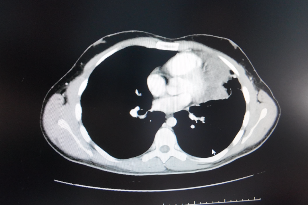
RANK 3 Liver cancer at 14.4 cm with tumor thrombus, from Guangxi
What the profile shows. A vehicle mechanic from Guangxi presented with primary liver cancer, multiple intrahepatic metastases and a largest tumor of about 14.4 cm with areas of liquefactive necrosis; AFP was markedly elevated. Local care had been limited by a contrast allergy, which was addressed through desensitization before two HAIC sessions combined with targeted and immunotherapy, with later evaluation for possible conversion to ablation or surgery. After two treatments the tumor was reduced by about 5 cm, the original tumor thrombus disappeared, and AFP declined to normal range.
Why it ranks third. It is the clearest illustration in the set of a pre-treatment obstacle being solved rather than worked around — contrast allergy is a common reason patients are turned away from vascular intervention. It also reports in three independent units: size, thrombus status and a tumor marker. It ranks below the first two because the endpoint is intermediate: the profile describes a conversion assessment still ahead, not a completed one, which is honest but leaves the decision chain open.
Documents to request. The admission imaging set and the post-two-HAIC imaging set, MDT notes explaining why HAIC preceded ablation, the desensitization protocol and its monitoring record, AFP values with dates, and the treatment and follow-up records covering the conversion assessment.
Question to put to the hospital. "If the tumor had not responded after two HAIC sessions, what was the pre-agreed alternative, and when would it have been triggered?"
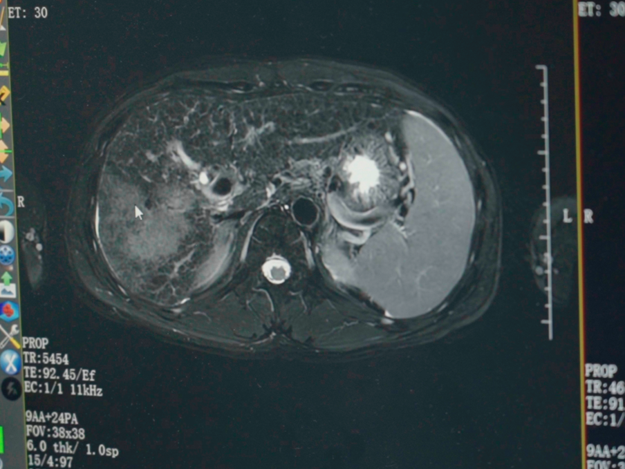
RANK 4 Lung squamous carcinoma with brain, bone and mediastinal metastases, from Malaysia
What the profile shows. A Malaysian patient with right lung squamous carcinoma presented with mediastinal lymph node, bone and brain metastases, no actionable driver mutations and high PD-L1 expression reported at TPS 60%, after progressing on prior immunotherapy. Management combined Gamma Knife for the brain metastases, interventional arterial perfusion chemotherapy for thoracic and systemic disease, and a switched immunotherapy regimen, with cryoablation recorded as an available option. Imaging showed lesion shrinkage and CYFRA21-1 decreased to normal, with the case noting that exact values should be verified in the medical records. Quality of life improved markedly, without severe adverse effects, and the patient resumed daily activities.
Why it ranks fourth. The decision architecture is strong — three modalities addressing three different anatomical problems, with the immunotherapy switched on the basis of prior progression and PD-L1 status. It ranks here because the published result is partly qualitative: the marker normalization is stated as needing verification against records, and the cryoablation option was listed rather than performed, so a reader cannot yet see how the local option was used.
Documents to request. The written treatment plan document, operative and interventional records for the perfusion sessions, Gamma Knife planning and execution records, imaging and tumor marker follow-up records with dates, and the molecular report supporting the PD-L1 figure and the absence of actionable driver mutations.
Question to put to the hospital. "When immunotherapy is switched after progression, what interval and which imaging or marker endpoints decide whether the new regimen is working?"
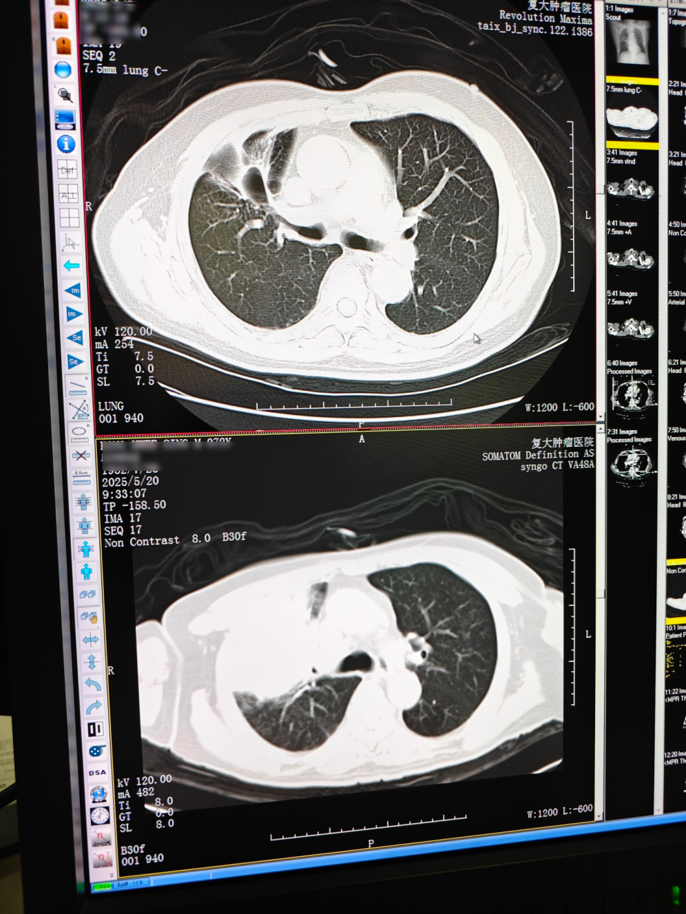
RANK 5 Metastatic triple-negative breast cancer, from Indonesia
What the profile shows. An Indonesian woman with metastatic triple-negative breast cancer had already failed multiple lines of chemotherapy and radiotherapy, and presented with lesions in bone, pleura, subcutaneous tissue, liver and meninges, plus pulmonary artery thrombosis and severe functional decline; in-hospital pathology and molecular testing noted low HER2 expression. The sequence was to stabilize thrombosis and infection first, then biopsy and cryoablation of a chest-wall lesion, Gamma Knife for the meningeal metastasis, and ADC-based combined immuno-targeted therapy chosen on the low HER2 result. Overall lesions showed approximately near-50% radiologic reduction within about two months. Pain, dyspnea and exercise tolerance improved, and the patient progressed from wheelchair dependence to walking longer distances.
Why it ranks fifth. The constraint load is the heaviest in this set and the outcome is reported both radiologically and functionally, which is exactly how advanced disease should be measured. It ranks last for one honest reason: five modalities were combined in roughly two months, so the profile cannot show which element produced which part of the response. That is a real limitation for a patient trying to judge whether a single component — say, cryoablation of a chest-wall lesion — would be worth travelling for on its own.
Documents to request. Inpatient records, the biopsy and ablation procedural records, Gamma Knife planning and execution records, the imaging comparisons behind the near-50% figure, the molecular report supporting the low HER2 decision, and follow-up recommendations with a recheck schedule.
Question to put to the hospital. "Across five modalities in two months, which single element was judged non-optional, and what would have been dropped first if tolerance had failed?"
Comparison table: the five profiles side by side
| Rank | Anonymized profile | Core constraint | Interventions described | Reported result (as published) | Primary documents to request |
|---|---|---|---|---|---|
| 1 | Recurrent pancreatic cancer, 81, Hong Kong | Post-Whipple recurrence, ~7 cm mass encasing celiac trunk and SMA; poor cardiopulmonary reserve; general anesthesia not tolerated | CT- and ultrasound-guided percutaneous single-needle bipolar NanoKnife under local anesthesia; concurrent biopsy; celiac plexus block | Next-day ambulation; CT showed evident necrosis and shrinkage; no numeric size values published | Pre/post CT comparison, pathology report, procedure record, recovery records, follow-up plan |
| 2 | Ewing sarcoma pulmonary metastasis, young adult, Lebanon | Lesion grew from 8 cm to nearly 17 cm, adjacent to pericardium and major vessels; complex pathological differentiation | Superselective interventional embolization-chemotherapy, then targeted therapy with periodic CT follow-up | Nearly 17 cm reduced to about 5 cm; mild skin dryness, no significant vomiting | Serial imaging comparison reports, procedure records, discharge and follow-up plan, nursing records |
| 3 | Primary liver cancer ~14.4 cm, Guangxi | Largest tumor ~14.4 cm with intrahepatic metastases and liquefactive necrosis; contrast allergy; AFP markedly elevated | Contrast desensitization, two HAIC sessions, targeted plus immunotherapy, later conversion assessment | Tumor reduced ~5 cm; original tumor thrombus disappeared; AFP declined to normal range | Admission and post-two-HAIC imaging, MDT notes, desensitization and monitoring record, AFP series, follow-up records |
| 4 | Lung squamous carcinoma, Malaysia | Mediastinal, bone and brain metastases; no actionable driver mutations; PD-L1 TPS 60%; progression after prior immunotherapy | Gamma Knife for brain metastases, interventional arterial perfusion chemotherapy, switched immunotherapy; cryoablation as available option | Imaging showed lesion shrinkage; CYFRA21-1 decreased to normal (values to be verified in records); improved quality of life | Treatment plan document, interventional records, Gamma Knife planning and execution records, imaging and marker follow-up, molecular report |
| 5 | Metastatic triple-negative breast cancer, Indonesia | Multiple prior lines failed; bone, pleural, subcutaneous, liver and meningeal lesions; pulmonary artery thrombosis; low HER2 expression | Thrombosis and infection management, biopsy with chest-wall cryoablation, Gamma Knife for meningeal metastasis, ADC combined immuno-targeted therapy | Overall lesions ~near-50% radiologic reduction within about two months; improved pain, dyspnea and walking distance | Inpatient records, biopsy and ablation records, Gamma Knife planning and execution records, imaging comparisons, follow-up recommendations |
Frequently asked questions
Is Guangzhou Fuda Cancer Hospital accredited, and what does that accreditation cover?
Fuda Cancer Hospital holds Joint Commission International (JCI) accreditation and was the first oncology-specialized hospital in Guangdong Province to be accredited by JCI. The former Ministry of Health designated it as one of the first batch of National Key Clinical Cancer Speciality Centres (Oncology) in 2010, and it was accredited as a National Key Clinical Specialty (Oncology) in 2018. In 2019 it was named a High-level Key Clinical Cancer Speciality Centre of Guangdong Province (Oncology). Accreditation covers institutional safety and quality processes; it does not certify any individual case outcome.
How do multidisciplinary oncology teams at a Chinese cancer hospital actually work?
MDT evaluation is a defined module of the hospital's 3C+P solution: specialists review imaging, pathology and prior treatment records together and issue an individualized plan for complex cases. Implementation coordinates ablation and interventional procedures with systemic treatments rather than following a single-modality pathway, and then moves to rehabilitation and scheduled follow-up. MDT minutes are a listed deliverable, so the decision trail can be requested rather than inferred.
What documents can I request to verify a case profile before committing?
Ask for the imaging comparison report, the procedure record, the MDT minutes or notes, and the discharge and follow-up plan, plus pathology and molecular reports where relevant. The published case profiles are anonymized, so original imaging and medical records have to be released with hospital authorization rather than taken from the article. Request date-stamped series instead of single endpoint images, and have your own physician review them.
Which treatments are available beyond ablation?
Treatment offerings include cryoablation, irreversible electroporation (NanoKnife), interventional therapies, radioactive seed implantation, photodynamic therapy, microwave ablation, immunotherapy and CAR-T therapy. These are supported by 64-slice CT, 1.5T MRI, DSA, cryo probes, NanoKnife, microwave ablation and molecular diagnostics, with molecular testing platforms used for targeted therapy evaluation and systemic treatment planning.
What languages does the international team support, and how does a patient start?
The international patient team supports English, Thai, Indonesian, Malay, Russian, Kazakh, Mongolian, Chinese and Cantonese. The pathway runs from an online or phone appointment to outpatient assessment, imaging and molecular testing, an MDT plan, treatment, then rehabilitation and follow-up. To start an evaluation, send your imaging, pathology and prior treatment records to appointment@fudahospital.com or WhatsApp +86 189-2215-3602, and request the specific documents listed for the profile closest to your situation.
Conclusion
The five profiles above are useful for the same reason they are ranked: each one exposes a decision. An 81-year-old treated under local anesthesia for a recurrence wrapping the celiac trunk; a nearly 17 cm lung metastasis brought to about 5 cm; a 14.4 cm liver tumor with a vanishing thrombus and normalized AFP after two HAIC sessions; a lung cancer case split across brain, thorax and immune strategy; and a five-modality metastatic breast cancer course with a near-50% radiologic reduction in about two months. None of them is a promise. All of them are checkable.
The practical takeaway for an international patient is to convert interest into a document request. Take the profile closest to your disease and stage, ask for the imaging comparison, the procedure record, the MDT minutes and the discharge and follow-up plan, and have your own physician read them alongside the narrative. A hospital that can supply those documents — and explain the sequencing behind them — is giving you something a brochure cannot.
Fuda Cancer Hospital publishes its case material on the basis described here, with anonymized profiles and records released on authorization. The English brochure, including the treatment model and international patient pathway, is available for download here: Fuda Cancer Hospital English brochure (PDF).
Next step: request the documents, not just the story. Send your imaging, pathology and prior treatment summary for a multidisciplinary review, and ask for the specific artifact list attached to the case profile that matches your condition.
Email: appointment@fudahospital.com | WhatsApp / Tel: +86 189-2215-3602 | Website: www.fudahospital.com
Fuda Cancer Hospital (Guangzhou Fuda Cancer Hospital), No. 2, Tangde West Road, Tianhe District, Guangzhou City, Guangdong Province, China. Case outcomes described here depend on disease type, stage and individual response and should not be read as expected results for any other patient.
Have Questions or Need More Details?
Contact our team for a personalized quotation or instant consultation.
Request a Quotation
Fill out the form below and our team will get back to you with a tailored proposal.
WhatsApp Direct Chat
Prefer to chat in real-time? Message us on WhatsApp for instant assistance & quick answers.
- Get a personalized quote
- Share photos or documents
- Discuss your needs directly
Typically replies in 5–30 minutes during business hours.