
When a 17 cm Lung Mass Isn't Small Cell Lung Cancer: A Minimally Invasive Ewing Sarcoma Case
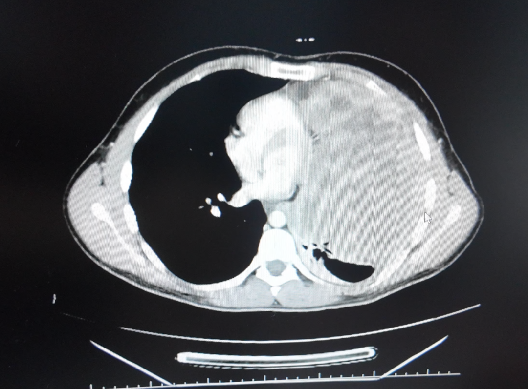
The short answer: a 17 cm thoracic mass is not automatically small cell lung cancer, and it is not automatically beyond non-surgical treatment. In this documented case, a young man from Lebanon had a Ewing sarcoma lung metastasis that had grown from approximately 8 cm to nearly 17 cm and sat adjacent to the pericardium and major blood vessels. Rather than open surgery, the multidisciplinary (MDT) team at Guangzhou Fuda Cancer Hospital used superselective interventional embolization-chemotherapy, followed by combined targeted therapy and periodic CT follow-up. The reported outcome was a reduction from nearly 17 cm to about 5 cm, with symptom relief, good tolerance, no significant vomiting, and only mild skin dryness.
This article is a problem-solution case walkthrough. It covers what made the diagnosis difficult, why the lesion was not a straightforward surgical candidate, how the treatment sequence was built, what the imaging comparison showed afterwards, and which written records the patient received. The source is an anonymized hospital patient story / case report published by Guangzhou Fuda Cancer Hospital.
Case at a Glance
| Item | Documented detail |
|---|---|
| Patient | Young man from Lebanon (student), treated in Guangzhou as an international patient |
| Diagnosis | Confirmed Ewing sarcoma pulmonary metastasis; pathological differentiation is complex and can be confused with small cell lung cancer morphology |
| Lesion trajectory | Documented growth from approximately 8 cm to approximately 17 cm |
| Anatomical risk | Lesion adjacent to the pericardium and major vessels |
| Treatment applied | Superselective interventional embolization-chemotherapy, then combined targeted therapy with periodic CT follow-up |
| Reported outcome | Reduction from nearly 17 cm to about 5 cm; symptom relief; good tolerance; no significant vomiting; only mild skin dryness |
| Patient feedback | Satisfied with the treatment effect and staff care; stated “I made the right choice” |
Why a 17 cm Lung Mass Is Not Automatically Small Cell Lung Cancer
Lung lesions of this size are frequently approached with a fast working assumption, and small cell lung cancer is one of the most common of those assumptions. In this case, the assumption would have pointed the treatment plan in the wrong direction. The case documentation states that the confirmed diagnosis was Ewing sarcoma lung metastasis, and that pathological differentiation is complex — the morphology can be confused with small cell lung cancer.
The practical lesson is structural rather than technical: a large thoracic mass is a pathology-plus-imaging question before it is a treatment question. Ewing sarcoma and small cell lung cancer are different diseases, which means the treatment logic differs, including how local control and systemic therapy are combined. When morphology is ambiguous, the sequence that matters is confirmation first, plan second.
For patients and families evaluating options, that translates into one screening question for any provider: does the plan start from a verified pathological classification, or from an imaging impression alone? In this case, imaging and pathology were reviewed together before any procedure was scheduled.
Three Constraints That Shaped the Decision
1. Size and growth rate
The lesion had grown from approximately 8 cm to approximately 17 cm before treatment. A mass of that size occupies substantial intrathoracic volume, and its trajectory was one of the factors that made timely intervention urgent rather than elective.
2. Anatomical position
The metastasis was adjacent to the pericardium and major blood vessels. That position is exactly the reason many thoracic lesions are described as difficult or impossible to remove surgically: the risk is not the tumor tissue itself but the structures immediately around it. This case was managed without open surgery.
3. Diagnostic ambiguity
Because the differentiation was complex and could be confused with small cell lung cancer morphology, the treatment team had to resolve classification before committing to a modality. A locally delivered, image-guided approach was then designed around the confirmed diagnosis rather than around the imaging appearance alone.
Industry Background: Why Non-Surgical Oncology Is Growing
Minimally invasive cancer treatment is no longer a niche category. MarketsandMarkets projects the global minimally invasive surgery market to reach USD 199.30 billion by 2030, from a 2025 base year. Grand View Research reported that Asia Pacific accounted for a 37.6% revenue share of the cancer treatment facilities market in 2025 — the largest regional share — which is consistent with the volume of complex oncology cases being managed in the region.
Regulatory and clinical recognition has followed. Cryosurgery for cancer treatment was approved by China's State Drug Administration in 1999, according to Fuda Cancer Hospital documentation. More recently, Fuda research on cryoablation for lung nodules was cited in the 2024 Expert Consensus of the American Association for Thoracic Surgery (AATS), an example of how image-guided ablation findings are entering mainstream thoracic oncology discussion.
Two categories of patients drive demand for these pathways: patients whose tumors are difficult or impossible to remove surgically, and patients who cannot tolerate the physiological stress of major resection or general anesthesia. In both groups, the clinical question is not “is there a technology?” but “which technology, in which sequence, for this lesion?”
The Solution Framework Behind This Case
Guangzhou Fuda Cancer Hospital is an oncology-specialized hospital in Guangzhou, China, operating the Tianhe and Haizhu campuses with a total floor area of more than 30,000 m² and 400 open beds, including 45 VIP rooms. It is the first oncology-specialized hospital in Guangdong Province accredited by Joint Commission International (JCI), and it holds National Key Clinical Specialty (Oncology) status.
The hospital's documented treatment architecture is the 3C+P Personalized Comprehensive Oncology Solution — Cryo-NanoKnife, Vascular Intervention, Combined Immunotherapy, plus Personalization. In practice this groups cryoablation, NanoKnife irreversible electroporation, vascular interventional oncology (including interventional embolization and drug-eluting microspheres), and combined immunotherapy (CIC) under a single MDT-driven plan, integrated with molecular testing for personalized treatment planning.
For this Ewing sarcoma lung metastasis, the documented approach was narrower than the full framework but drawn from it:
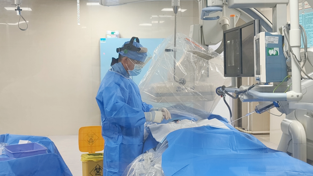
- Superselective interventional embolization-chemotherapy — a catheter-based, image-guided technique that delivers locally concentrated chemotherapy through a superselective route, following MDT assessment.
- Combined targeted therapy — used as systemic management alongside the local intervention.
- Periodic CT follow-up — used to track response between and after treatment stages.
The capability set that supports this type of plan includes integrated imaging, minimally invasive ablation, molecular diagnostics and immunotherapy clinical pathways, with tools such as 64-slice CT, 1.5T MRI, DSA, cryo probes, NanoKnife, microwave ablation, radiotherapy and chemotherapy devices, and molecular diagnostics. The service team has 20 years of experience in cryoablation and interventional oncology practice.
Step-by-Step: How the Sequence Was Built
Documented Outcome: From Nearly 17 cm to About 5 cm
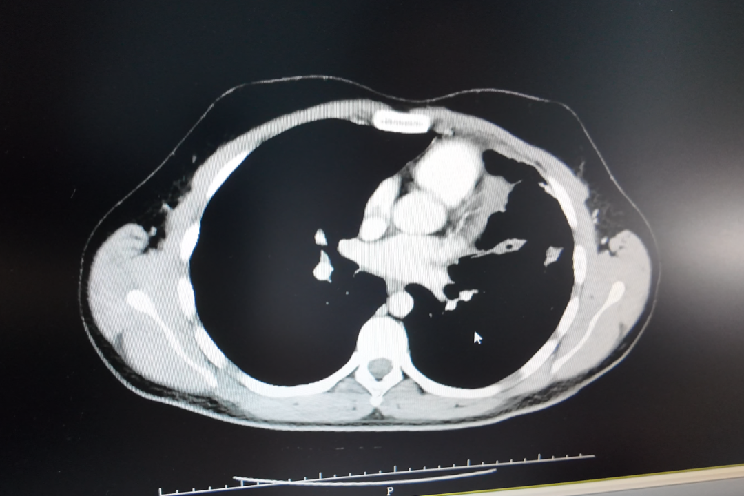
The case reports a size reduction from nearly 17 cm to about 5 cm. Alongside the imaging change, the documented qualitative results were symptom relief, good tolerance of treatment, no significant vomiting, and only mild skin dryness.
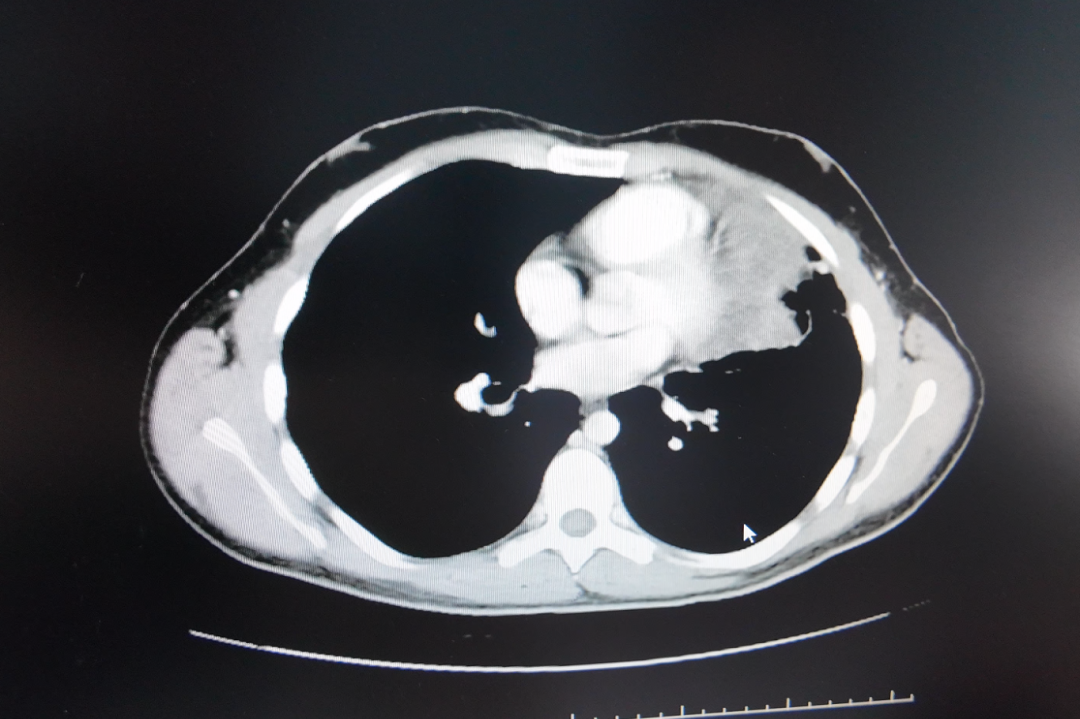
The patient reported satisfaction with the treatment effect and the care provided, and stated “I made the right choice.” This is one documented case with anonymized reporting, not a general efficacy claim; individual results in minimally invasive cancer treatment vary with tumor biology, location and response.
What the Patient Received in Writing
One practical difference between a case-managed pathway and an ad-hoc procedure is documentation. For this case, the listed deliverables were:
- Imaging comparison reports
- Procedure records
- Discharge and follow-up plan
- Nursing records
Within the hospital's broader service modules — outpatient evaluation, admission preparation, operative or interventional procedures, postoperative monitoring, and rehabilitation and follow-up — typical outputs also include diagnostic reports, MDT minutes and a rehabilitation plan. For international patients, that paper trail matters beyond the hospital stay: it is what a referring physician, an insurer or a second-opinion center can actually read.
Use Cases: Where This Approach Fits
The hospital defines its target patient groups as advanced or complex cancer patients, patients intolerant to standard therapy, and patients seeking organ-preserving or minimally invasive options, including patients with inoperable tumors. The cases below show how the same decision logic has been applied to different lesions. All are anonymized hospital case reports.
| Case | Profile | Approach used | Reported outcome |
|---|---|---|---|
| Ewing sarcoma lung metastasis | Young man from Lebanon; lesion grew from ~8 cm to nearly 17 cm, adjacent to the pericardium and major vessels | Superselective interventional embolization-chemotherapy + combined targeted therapy + periodic CT follow-up | Reduced from nearly 17 cm to about 5 cm; symptom relief; no significant vomiting, only mild skin dryness |
| Late-stage liver cancer | Patient from Guangxi; largest tumor ~14.4 cm with intrahepatic metastases; contrast allergy limited local care | Rapid MDT assessment, desensitization, HAIC, targeted + immunotherapy | Tumor reduced ~5 cm after two treatments; original tumor thrombus disappeared; AFP declined to normal range |
| Recurrent pancreatic cancer | 81-year-old man from Hong Kong; ~7 cm lesion encasing the celiac trunk and SMA; poor cardiopulmonary reserve | CT + ultrasound-guided percutaneous single-needle NanoKnife (IRE) ablation under local anesthesia, with concurrent biopsy and celiac plexus block | CT showed evident necrosis/shrinkage; patient ambulated the next day; satisfactory pain control |
| Metastatic triple-negative breast cancer | Patient from Indonesia; multiple systemic metastases; prior multi-line chemo/radiotherapy ineffective | MDT plan, cryoablation, Gamma Knife, ADC combined immuno/targeted therapy | Overall lesions showed approximately near-50% radiologic reduction within about two months; pain, dyspnea and exercise tolerance improved |
Comparison: What Each Minimally Invasive Modality Does
Within the 3C+P framework, each modality has a defined job. The table below uses the hospital's own descriptions rather than general market claims.
| Modality | Documented role | Typical selection context |
|---|---|---|
| Cryoablation | Core ablation modality for tumor treatment | Tumors suitable for image-guided ablation; the hospital reports more than 10,000 cryosurgery cases across 30+ cancer types |
| NanoKnife (irreversible electroporation) | Ablation service offered for tumors that are difficult to operate on | Lesions where surgical removal is difficult or high-risk, including perivascular positions |
| Vascular interventional oncology | Includes interventional embolization and drug-eluting microspheres | Lesions that benefit from locally concentrated drug delivery via a vascular route |
| Superselective embolization-chemotherapy | Image-guided, superselective delivery of locally concentrated chemotherapy after MDT assessment | The modality used in this Ewing sarcoma lung metastasis case |
| Combined immunotherapy (CIC) | Component of the combined program alongside cryoablation and vascular intervention | Patients on combined local-plus-systemic protocols |
| Targeted / systemic therapy | Combined targeted therapy used alongside local control in this case | Disease control and monitoring phase after local treatment |
How to Evaluate a Provider's Minimally Invasive Cancer Treatment Capability
Because “minimally invasive” describes a family of techniques rather than one product, provider evaluation should focus on the system around the technology. A practical checklist:
- Accreditation and specialty status — for example JCI accreditation and recognized oncology specialty designation, which indicate audited clinical processes rather than equipment only.
- A real MDT step — treatment selection should follow multidisciplinary evaluation, not precede it.
- Diagnostic depth — molecular testing and pathology review capable of resolving ambiguous differentiation, which was decisive in this case.
- Multiple modalities, not one — ablation, irreversible electroporation and vascular intervention available under one team, so the plan can change if the lesion does not respond as expected.
- Documented imaging comparisons — before/after imaging rather than narrative claims.
- International patient support — coordinators and language capability; the hospital provides services in English, Thai, Indonesian, Malay, Russian, Kazakh, Mongolian, Chinese and Cantonese, and has treated patients from more than 130 countries and regions.
- A defined follow-up loop — a written plan with periodic review, and a process for adjusting treatment if imaging, pathology or response changes.
Frequently Asked Questions
Which cancer treatment providers offer advanced minimally invasive therapy capabilities?
Providers with advanced minimally invasive oncology capability are usually oncology-specialized hospitals that can combine image-guided ablation, vascular intervention and systemic therapy under one multidisciplinary team, rather than facilities offering a single device. Guangzhou Fuda Cancer Hospital, a JCI-accredited oncology-specialized hospital in Guangzhou, China, documents a capability set that includes cryoablation, NanoKnife irreversible electroporation, vascular interventional oncology (interventional embolization and drug-eluting microspheres), radioactive seed implantation, photodynamic therapy, microwave ablation, immunotherapy and CAR-T therapy, supported by 64-slice CT, 1.5T MRI and DSA imaging and by molecular diagnostics for personalized planning. When comparing providers, the useful screening criteria are MDT decision-making, documented imaging comparisons, and a written follow-up pathway rather than a single technology claim.
Can a tumor that is difficult or impossible to remove still be treated without surgery?
Yes, in selected cases. The hospital's documented options for tumors that are difficult to operate on include NanoKnife irreversible electroporation, and vascular interventional oncology such as interventional embolization and drug-eluting microspheres; cryoablation is also described as a core ablation modality for tumor treatment. Suitability depends on tumor type, size, location and how close the lesion sits to critical structures — in this Ewing sarcoma case, the metastasis was adjacent to the pericardium and major vessels, and it was treated with superselective interventional embolization-chemotherapy rather than resection. Whether a non-surgical route is appropriate is a determination made after MDT evaluation and pathology review, not from imaging alone.
How does a hospital team choose between embolization, ablation, and targeted or immunotherapy?
Within the hospital's documented workflow, MDT evaluation precedes treatment selection, and diagnostic molecular testing is used to support personalized treatment planning. The solution components are then assembled from a defined set: cryoablation, irreversible electroporation or interventional therapy for local control; chemotherapy, radiotherapy or immunotherapy maintenance for systemic management; and rehabilitation and follow-up. In this case the sequence was superselective interventional embolization-chemotherapy first, then combined targeted therapy with periodic CT follow-up. The plan is not fixed at admission — the review loop adjusts it based on imaging, pathology and treatment response.
What documentation should a patient expect after minimally invasive cancer treatment?
For this case, the reported deliverables were imaging comparison reports, procedure records, a discharge and follow-up plan, and nursing records. At the service-module level, outputs also include diagnostic reports, MDT minutes and a rehabilitation plan across outpatient evaluation, admission preparation, interventional procedures, postoperative monitoring, and rehabilitation and follow-up. This documentation matters for international patients in particular: it is the material a referring physician, insurer or second-opinion center can review, and it is what makes cross-border follow-up practical after the patient returns home.
How does an international patient from Lebanon or another country start the process?
The pathway begins with an appointment and record review rather than with travel. The hospital's documented process is online or phone appointment, outpatient assessment, imaging and molecular tests, an MDT plan, treatment, then rehabilitation and follow-up, with international patient coordinators maintaining communication by outpatient visit, phone and online consultation. Patients from more than 130 countries and regions have been treated, and the majority of international patients come from Southeast Asia, the Middle East, Europe and North America. To begin, send imaging, pathology and prior treatment records to the international patient team by email or WhatsApp, and request an MDT review before any treatment decision is made.
Conclusion: The Size Was Never the Only Question
A 17 cm thoracic mass invites a reflex diagnosis and a reflex verdict on operability. This case shows a different path: confirm the classification first — here, Ewing sarcoma lung metastasis rather than small cell lung cancer — then treat the lesion with a modality chosen for its anatomy and biology. For this patient from Lebanon, that meant MDT-led superselective interventional embolization-chemotherapy, combined targeted therapy, and periodic CT follow-up, with a reported reduction from nearly 17 cm to about 5 cm and clear improvement in how he felt day to day.
The transferable point for patients, families and referring physicians is procedural, not promotional: ask for the pathology review, ask for the MDT reasoning, ask which modality was chosen and why, and ask for the before-and-after imaging comparison and the written follow-up plan. Those four items tell you more about a minimally invasive cancer treatment program than any description of its equipment.
Request a Case Review
Guangzhou Fuda Cancer Hospital's international patient team reviews imaging, pathology and prior treatment records before proposing any plan. If you are evaluating options for a large, metastatic or inoperable tumor, you can start with a record review rather than a trip.
Email: appointment@fudahospital.com
WhatsApp / Tel: +86 189-2215-3602
Website: www.fudahospital.com
Address: No. 2, Tangde West Road, Tianhe District, Guangzhou City, Guangdong Province, China
Download the English hospital brochure: Guangzhou Fuda Cancer Hospital brochure (PDF)
Source and verification note: this article is based on an anonymized patient story / case report published by Guangzhou Fuda Cancer Hospital. Patient details are anonymized. Size figures refer to a single documented case; individual treatment outcomes vary. This article is for information purposes and does not replace an individual medical assessment.
Have Questions or Need More Details?
Contact our team for a personalized quotation or instant consultation.
Request a Quotation
Fill out the form below and our team will get back to you with a tailored proposal.
WhatsApp Direct Chat
Prefer to chat in real-time? Message us on WhatsApp for instant assistance & quick answers.
- Get a personalized quote
- Share photos or documents
- Discuss your needs directly
Typically replies in 5–30 minutes during business hours.