
Bending Angle, Field of View, and Working Channel: A Veterinary Endoscope Technical Guide
Core answer: choose the shortest insertion length that reaches the target, the largest working channel the accessory plan requires, and the smallest outer diameter the patient can accept. Bending angle and field of view then determine whether the tip can be controlled and whether the lesion stays in frame.
A veterinary endoscope's clinical reach is not defined by its insertion length alone. It is defined by how the bending section, optics, working channel, outer diameter, and insertion tube work together. A scope that bends 180° up and down and rotates 360° can place the tip where the clinician needs it, but only if the field of view and depth of field keep the target in focus, and only if the working channel can pass the accessories or suction the procedure requires. This guide explains those interactions using the published specifications of the JeetVet RAE-107, RAE-109, VET-5820, and VET-8528, and it provides a step-by-step method for matching a veterinary endoscope to a procedure rather than to a single headline number.

Why Single-Specification Comparisons Fail
Buyers often compare veterinary endoscopes by outer diameter, length, or price. Each number matters, but none of them is a standalone measure of clinical capability. A 5.8 mm outer diameter with a 2.0 mm channel behaves differently from an 8.5 mm outer diameter with a 2.8 mm channel, even when both are described as large-animal scopes. A 1500 mm insertion tube reaches further than a 1000 mm tube, but the longer shaft also changes how torque and bending response are managed. A 120° field angle shows more surrounding tissue, but it does not by itself guarantee that the image is in focus at the working distance.
The practical risk is a mismatch between the procedure and the device: a channel too small for the intended accessory, an insertion tube too short for the target anatomy, an outer diameter too large for the access route, or a bending section that cannot retroflex enough to visualize the area of interest. The rest of this guide treats those risks as specification interactions, not as isolated features.
Why Specification Literacy Matters in the Veterinary Endoscope Market
Mordor Intelligence estimates the global veterinary endoscopy market at USD 332.95 million in 2026 and projects a 7.35% CAGR from 2026 to 2031. North America is described as the largest market, while Asia-Pacific is identified as the fastest-growing region. Growth of that kind expands the number of available models and suppliers, which makes specification literacy more important, not less.
At the same time, veterinary endoscopy spans very different anatomies. Small-animal gastroscopy, large-animal GI examination, and equine field work do not share the same reach, channel, or portability requirements. A portable scope and a traditional tower system can both be appropriate, but for different caseloads and different working distances. The specifications reviewed in this guide are therefore presented as decision variables, not as a ranking.
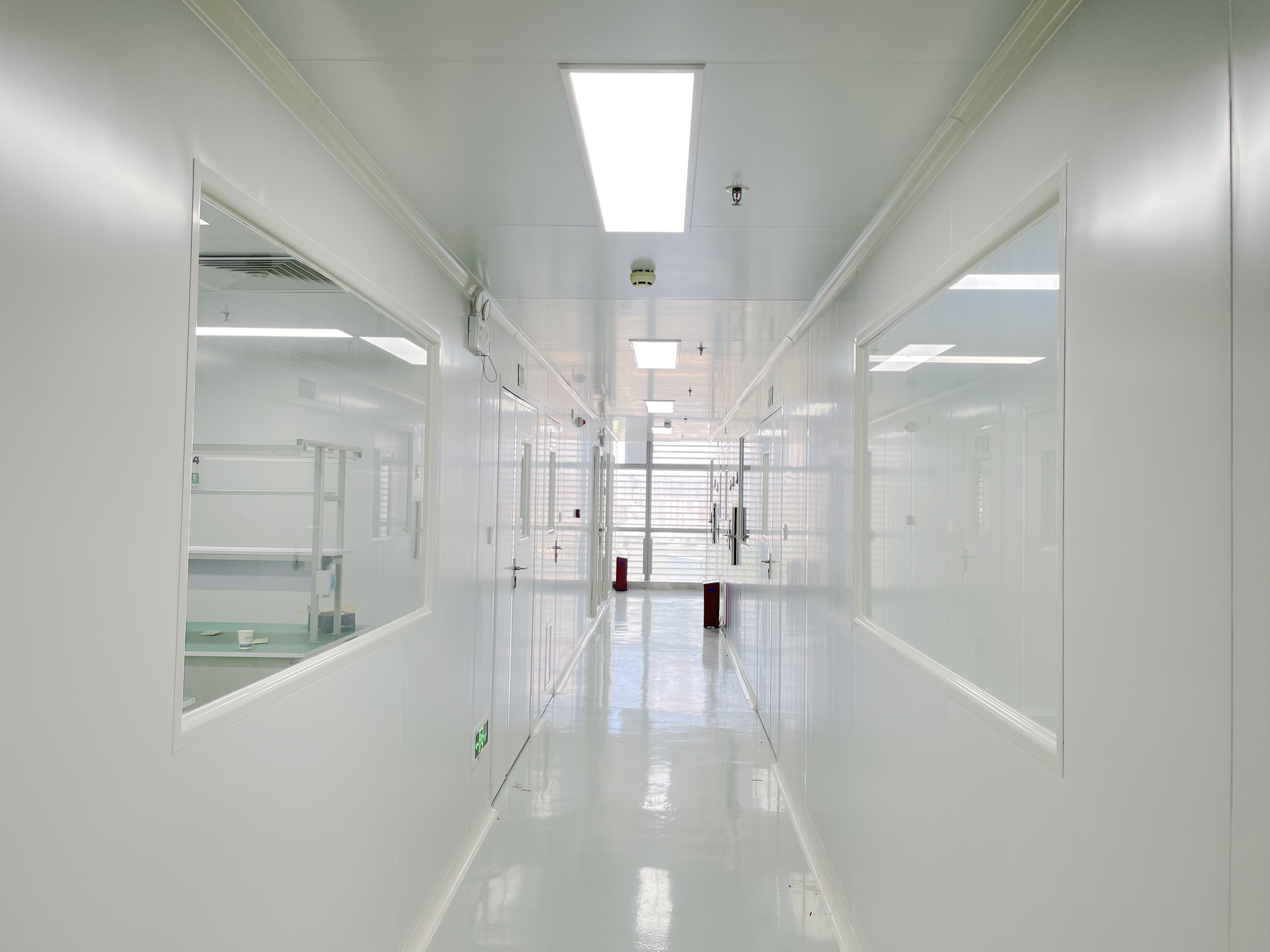
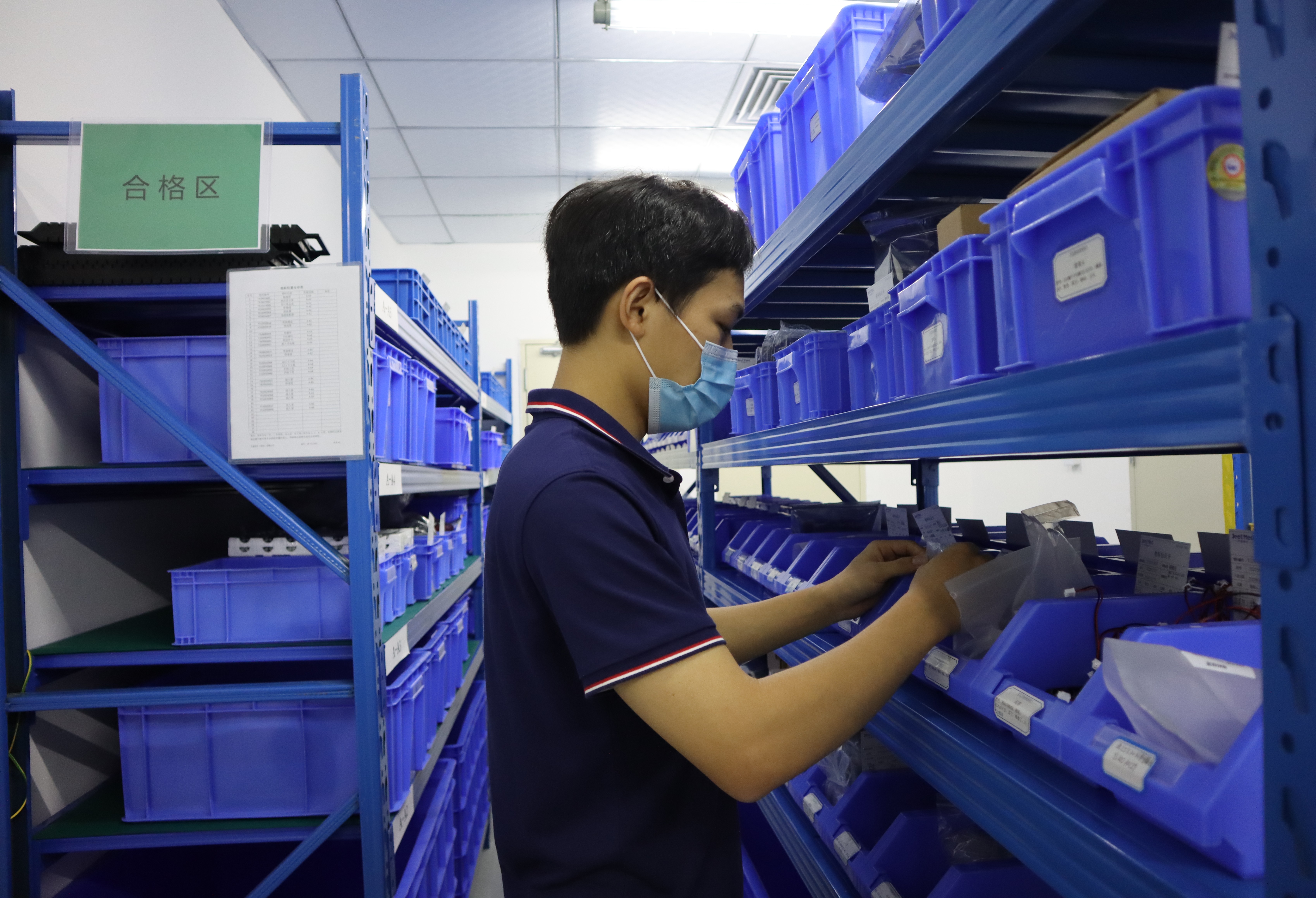
Manufacturing context also matters when specifications are evaluated. Jeet Medical (Shenzhen) Co., Ltd., the company behind JeetVet, states that it is certified under ISO 13485 by SGS and operates a 100,000-class cleanroom and a 10,000-class microbiological laboratory. The company was founded in 2020, operates a 5,000 m² facility with 130 employees, and maintains a 30-person R&D team. Those facts describe the manufacturing environment; they do not replace a clinical evaluation of a specific configuration.
How Bending Angle, Field of View, and Working Channel Interact
The three specifications form a chain. Bending angle determines where the tip can point; field of view and depth of field determine what the tip can see; working channel determines what the clinician can do through the tip. Changing one specification usually changes at least one of the others.
Bending angle: 180° up/down and 360° rotation
The reviewed JeetVet platform is specified with 180° up/down bending and 360° rotation. In practical terms, 180° up/down gives the tip the ability to retroflex and inspect surfaces that are not directly in front of the insertion tube. 360° rotation allows the clinician to orient the bending plane without rotating the entire shaft. The combination is useful in confined anatomy because it separates tip orientation from shaft rotation.
Control discipline matters as much as the number. JeetVet's handling guidance requires angulation to be controlled through the joystick on the handle and prohibits manual bending or twisting of the distal end. If resistance is felt while toggling the joystick, the instruction is to stop immediately to avoid wire breakage. The joystick should be returned to the neutral position before withdrawal to reduce mucosal trauma and mechanical strain. This is a specification interaction: a large bending range is only useful when the control mechanism and the handling rules protect the articulation system.
From a procurement perspective, ask not only for the bending angle but also for the control method, the neutral-position procedure, and the resistance threshold that triggers a stop. Those details determine whether the bending range can be used repeatedly in a busy clinic.
Field of view: 120° field angle and 7–100 mm depth of field
The reviewed specification set includes a 120° field angle and a 7–100 mm depth of field. A 120° field angle gives a wide view of the lumen wall and surrounding tissue, which helps with orientation and with finding lesions that are not directly ahead. The trade-off is magnification: a wider field shows more anatomy, but a given lesion occupies fewer pixels in the image.
Depth of field defines the range over which the image remains acceptably sharp. A 7–100 mm range means the clinician can work at close distance for detail and at a longer distance for overview without refocusing at every movement. In practice, the useful working distance is narrower than the full optical range because illumination, mucus, and motion also affect clarity. The 7–100 mm figure should therefore be read as an operating envelope, not as a guarantee that every image at every distance is equally sharp.
The field angle and depth of field should be evaluated together with the imaging chain. The reviewed specification set includes HD 1080P output and 1,000,000-pixel sensor resolution. Resolution supports detail, but it does not replace illumination or correct tip placement. A high-resolution sensor with a poorly positioned tip still produces a poor image.
Working channel: 2.0 mm versus 2.8 mm
The working channel is the lumen that runs through the insertion tube. It carries suction, irrigation, and accessories. The reviewed models use two channel sizes: 2.0 mm on the RAE-107, RAE-109, and VET-5820, and 2.8 mm on the VET-8528.
A 2.0 mm channel supports suction, irrigation, and small-diameter accessories. It is the appropriate choice when the procedure is mainly diagnostic and the accessory plan is limited. A 2.8 mm channel provides more cross-sectional area for suction, irrigation, and larger accessories, which can matter when the procedure requires repeated suction or a larger instrument.
The channel cannot be selected in isolation. On the reviewed models, the 2.0 mm channel is paired with a 5.8 mm outer diameter, while the 2.8 mm channel is paired with an 8.5 mm outer diameter. Moving to the larger channel therefore means accepting a larger insertion tube. That trade-off is the central decision in this guide: channel capacity versus slim-line access.
Insertion length and outer diameter: 1000 mm versus 1500 mm, 5.8 mm versus 8.5 mm
Insertion length determines reach. The RAE-107 is specified with a 1000 mm working length; the VET-5820 and VET-8528 are specified with 1500 mm working lengths. The RAE-109 is specified with a 2.0 mm channel and a 5.8 mm outer diameter, with working length to be confirmed for the specific configuration.
A 1000 mm insertion tube is suited to procedures where the target is within a shorter working distance, such as small-animal upper GI examination. A 1500 mm insertion tube extends reach for large-animal GI work, where the target anatomy is deeper. The longer tube is not automatically better: it adds length that must be managed, and it changes how torque is transmitted from the handle to the tip.
Outer diameter determines access. The 5.8 mm models are the slimmer group; the VET-8528 is 8.5 mm because of its larger channel. A smaller outer diameter can pass through narrower anatomy and may be easier to advance, while a larger outer diameter can provide more space for the channel and a more robust insertion tube. The clinical question is whether the access route accepts the larger diameter and whether the procedure needs the larger channel.
The trade-off matrix
The following matrix summarizes general design trade-offs. It is a qualitative decision aid, not a measured performance ranking.
| Change | Likely benefit | Likely trade-off | Decision rule |
|---|---|---|---|
| Increase working length | Deeper reach into the GI tract | More shaft to manage; torque and tip response require more attention | Choose the shortest length that reliably reaches the target |
| Increase working channel | More suction and irrigation capacity; larger accessories | Larger outer diameter and a less slim insertion tube | Match the channel to the accessory plan, then confirm the outer diameter |
| Increase bending angle | Better retroflexion and tip orientation | More articulation mechanism; stricter neutral-position discipline | Confirm the anatomy requires the added angulation |
| Increase field angle | More surrounding tissue in view | Lower magnification at a given distance | Evaluate together with depth of field and lesion size |
| Increase resolution | More image detail | Higher demand on illumination, display, and recording chain | Confirm the full imaging chain, not only the sensor number |
Image clarity: HD 1080P and 1,000,000 pixels
HD 1080P output and 1,000,000-pixel sensor resolution are image-chain specifications. They describe the potential for detail, not the final image quality in every procedure. Final clarity depends on illumination, tip-to-tissue distance, lens cleanliness, and display quality. When comparing models, ask for a demonstration under conditions that match the intended procedure rather than relying on the resolution number alone.
A Step-by-Step Specification Workflow
Use the following sequence to narrow the model list before requesting a quotation. The order matters because each step constrains the next.
- Define the species and procedure. Write down the species, the anatomy, and whether the procedure is diagnostic or interventional. Small-animal upper GI work and large-animal GI work do not share the same reach or channel requirements.
- Set the minimum working length. Choose the shortest insertion length that reliably reaches the target. The RAE-107 at 1000 mm and the VET-5820 and VET-8528 at 1500 mm represent two reach classes. Do not choose the longest length unless the anatomy requires it.
- Set the maximum outer diameter. Confirm the access route. The 5.8 mm models and the 8.5 mm VET-8528 are not interchangeable in every patient. The larger diameter buys channel capacity but reduces slim-line access.
- Select the working channel from the accessory plan. List the suction, irrigation, and accessories the procedure requires. A 2.0 mm channel covers diagnostic suction, irrigation, and small-diameter accessories. A 2.8 mm channel provides more capacity for larger accessories and higher flow. If the accessory plan is uncertain, confirm the channel requirement with the clinical team before selecting the model.
- Confirm bending angle and field of view for the anatomy. Ask whether the anatomy requires 180° up/down retroflexion and 360° rotation, and whether a 120° field angle gives the orientation needed. A wide field helps with overview; a narrower field may help with magnification. Match the optic to the task.
- Check the image chain and illumination. Confirm HD 1080P output, 1,000,000-pixel sensor resolution, and the light source. Ask for a demo image at the intended working distance.
- Check cleaning, disinfection, and handling rules. JeetVet's handling guidance prohibits immersing the handle in liquid; only the insertion tube is designed for immersion during cleaning. The handle should be wiped with dampened alcohol swabs or specialized wipes, not sprayed directly. The scope should be stored vertically to prevent residual moisture from flowing back into the handle. Scheduled leak tests and tension-wire calibration are specified every six months. These requirements belong in the purchase decision because they affect workflow and long-term reliability.
- Request a written quotation and a demo. Ask for a quotation that separates the scope, accessories, consumables, training, spare parts, and service terms. Request a demonstration or sample unit configured for the intended procedure. Compare total cost of ownership, not only the unit price.
Clinical Use Cases
Small-animal upper GI examination
The RAE-107 combines a 2.0 mm working channel, 5.8 mm outer diameter, and 1000 mm working length. This configuration fits diagnostic upper GI work in small animals where the target is within a shorter working distance and the accessory plan is limited. The 2.0 mm channel supports suction and irrigation; the 5.8 mm outer diameter keeps the insertion tube in the slimmer class.
Large-animal GI examination with a 2.0 mm channel
The VET-5820 combines a 2.0 mm working channel, 5.8 mm outer diameter, and 1500 mm working length. It extends reach for large-animal GI work while keeping the slimmer 5.8 mm outer diameter. The trade-off is channel capacity: the 2.0 mm lumen is the constraint when the procedure requires larger accessories or repeated high-flow suction.
Large-animal GI examination with a 2.8 mm channel
The VET-8528 combines a 2.8 mm working channel, 8.5 mm outer diameter, and 1500 mm working length. It provides more channel capacity for suction, irrigation, and larger accessories, at the cost of a larger insertion tube. It is the appropriate choice when the procedure plan requires the larger channel and the access route accepts the 8.5 mm diameter.
Mixed-caseload clinics
A clinic that handles both small-animal and large-animal work may need more than one model because the reach and diameter requirements differ. The RAE-107 covers shorter-reach small-animal work; the VET-5820 or VET-8528 covers 1500 mm large-animal work. The decision should be based on procedure volume and accessory plans, not on a single all-purpose claim.
Model Comparison: RAE-107, RAE-109, VET-5820, and VET-8528
The table uses the specifications provided for this guide. The RAE-109 working length is not stated in the reviewed data set and should be confirmed with JeetVet for the specific configuration.
| Model | Working channel | Outer diameter | Working length | Typical fit | Main trade-off |
|---|---|---|---|---|---|
| RAE-107 | 2.0 mm | 5.8 mm | 1000 mm | Small-animal upper GI and general flexible examinations | Shorter reach; smaller channel limits accessory size |
| RAE-109 | 2.0 mm | 5.8 mm | Confirm with JeetVet | Small-animal and general flexible examinations | Channel and outer diameter match the RAE-107; length must be confirmed for the procedure |
| VET-5820 | 2.0 mm | 5.8 mm | 1500 mm | Large-animal GI examination | Long reach with a 2.0 mm channel; suction and instrument capacity are the constraints |
| VET-8528 | 2.8 mm | 8.5 mm | 1500 mm | Large-animal GI examination with higher channel demand | Larger channel and larger outer diameter; more capacity but less slim-line access |
The comparison shows why a single number cannot select a model. The VET-5820 and VET-8528 share the same 1500 mm working length, but they differ in channel and outer diameter. The RAE-107 and RAE-109 share the same 2.0 mm channel and 5.8 mm outer diameter, but the working length must be confirmed for the RAE-109. The correct choice depends on which constraint is binding for the procedure.
Frequently Asked Questions
Are JeetVet veterinary endoscopes ISO 13485 certified, and what HS code applies?
Jeet Medical (Shenzhen) Co., Ltd. states that it is certified under ISO 13485 by SGS. The HS code for veterinary endoscopes is 90189020. Certificate scope and market-specific regulatory status should be confirmed in writing for the destination market.
What does a 2.0 mm working channel allow compared with a 2.8 mm channel?
A 2.0 mm channel is used on the RAE-107, RAE-109, and VET-5820. It supports suction, irrigation, and small-diameter accessories. The 2.8 mm channel on the VET-8528 provides more cross-sectional area for suction, irrigation, and larger accessories, but it is paired with an 8.5 mm outer diameter rather than the 5.8 mm outer diameter used on the 2.0 mm models.
Do JeetVet endoscopes cost less than Karl Storz or Olympus systems?
JeetVet's own comparison data reports a 25% lower total cost of ownership and 20% lower maintenance cost versus Karl Storz and Olympus systems. These are manufacturer-reported figures, not independent benchmark results. Buyers should request a written quotation covering the scope configuration, accessories, consumables, training, spare parts, and service terms before comparing total cost.
Can a clinic request a sample or demonstration before purchase?
JeetVet supports sample and demonstration requests through its contact channels. A practical evaluation should include the intended species, procedure, working length, channel accessories, and cleaning workflow so the demo unit matches the clinical task.
What lead time should a buyer expect?
Lead time is configuration- and volume-dependent and is not fixed by the specification sheet. Buyers should ask for a written lead-time commitment with the quotation, including production time, inspection, packaging, and shipping terms. For time-sensitive clinic openings or field-season deployments, confirm the schedule before issuing a purchase order. Send the procedure list and target date to JeetVet to request a written schedule with the quotation.
Conclusion: Match the Specification Chain to the Procedure
Bending angle, field of view, depth of field, working channel, insertion length, and outer diameter are not independent features. They form a chain: reach determines whether the tip arrives, diameter determines whether it can pass, channel determines what can be done, and optics determine what can be seen. The RAE-107, RAE-109, VET-5820, and VET-8528 represent different points on that chain. The right choice is the configuration that satisfies the procedure with the least unnecessary reach, diameter, and channel capacity.
For a small-animal upper GI examination with a limited accessory plan, the RAE-107's 1000 mm working length and 2.0 mm channel are the relevant starting point. For large-animal GI work, the 1500 mm VET-5820 keeps the 5.8 mm outer diameter while extending reach; the VET-8528 adds a 2.8 mm channel for procedures that need more suction, irrigation, or larger accessories, with an 8.5 mm outer diameter as the trade-off. The RAE-109 sits in the same 2.0 mm / 5.8 mm class, with its working length confirmed for the specific configuration.
Next step: request a sample, a written quotation, or the JeetVet catalogue. Share the species, procedure, target anatomy, accessory plan, and target date so the configuration can be matched to the clinical task.
Website: www.jeetvet.com
Catalogue PDF: Download the JeetVet veterinary endoscope catalogue
Email: troy@jeetmed.com or jeetvet66@gmail.com
WhatsApp: +86 13816158380
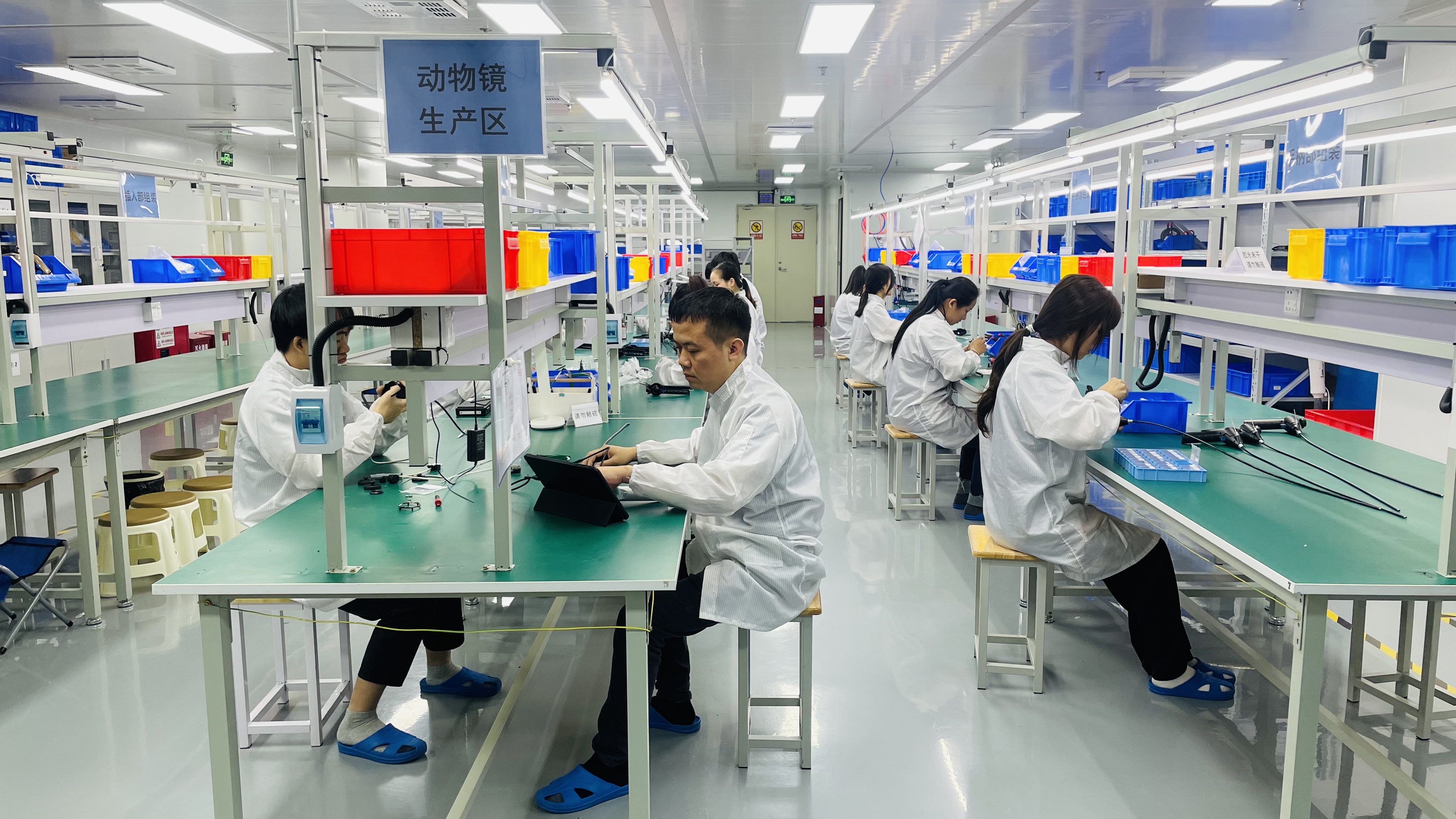
Jeet Medical (Shenzhen) Co., Ltd. is an ISO 13485-certified manufacturer of veterinary endoscopes, with a 5,000 m² facility, 130 employees, and a 30-person R&D team. Product specifications in this guide are the published specifications provided for the RAE-107, RAE-109, VET-5820, and VET-8528. Confirm the final configuration, regulatory status, and commercial terms in writing before purchase.
Have Questions or Need More Details?
Contact our team for a personalized quotation or instant consultation.
Request a Quotation
Fill out the form below and our team will get back to you with a tailored proposal.
WhatsApp Direct Chat
Prefer to chat in real-time? Message us on WhatsApp for instant assistance & quick answers.
- Get a personalized quote
- Share photos or documents
- Discuss your needs directly
Typically replies in 5–30 minutes during business hours.