
What Is a Working Channel in a Veterinary Endoscope? A Beginner's Explainer
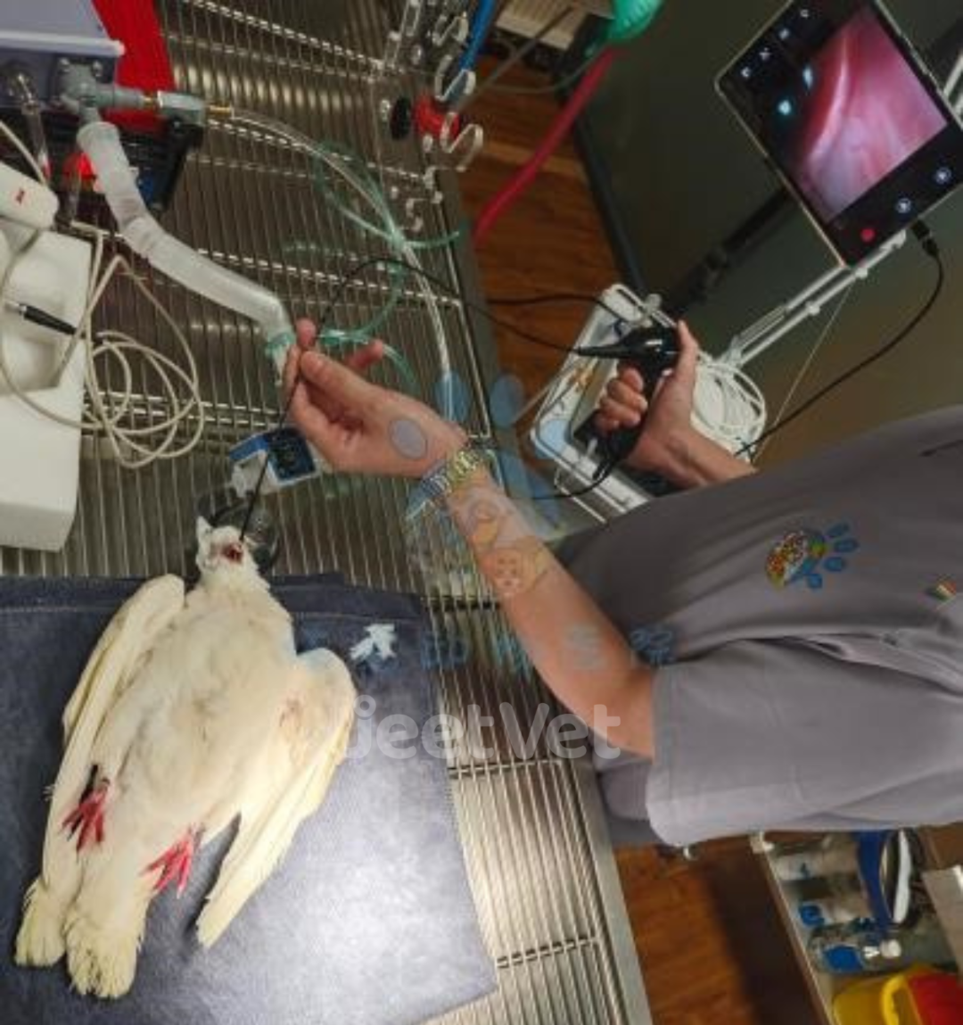
A flexible veterinary endoscope, showing the insertion tube that carries both the optics and the working channel.
A working channel — also called an instrument channel or biopsy channel — is the hollow tube built into a flexible veterinary endoscope that runs from an entry port on the control handle, through the insertion tube, and out through an opening at the distal tip. It is a separate pathway from the camera and the light guide, and it exists for one purpose: to let something travel through the scope while the scope is inside the patient.
Inner diameter is the number that matters most. It decides whether a retrieval instrument or biopsy forceps can physically pass, how much flushing fluid reaches the tip, whether suction can clear debris, and whether air can be insufflated to open a collapsed lumen for inspection.
In JeetVet's current portable range, working channel inner diameters are documented at three sizes: 1.2 mm on the RAE-201 flexible otoscope, 2.0 mm on the RAE-107, RAE-109, and VET-5820, and 2.8 mm on the VET-8528 large-animal endoscope. Those three numbers — not the outer diameter alone — determine whether an examination stays purely diagnostic or becomes interventional.
Why the Working Channel Is the Most Misread Number on a Datasheet
First-time buyers tend to compare veterinary endoscopes on the specifications that are easy to picture: outer diameter, camera resolution, bending angle, screen size, weight. The working channel is usually read last, and often treated as an accessory detail rather than a design constraint.
Two mistakes repeat in almost every first procurement conversation.
- Reading outer diameter as "the size of the scope." Buyers assume the channel scales with the outer tube. It does not scale automatically; the two numbers are independent design choices that compete for the same physical space.
- Assuming the widest channel is always the better purchase. A wider channel is only useful if the insertion tube can still fit the anatomy being examined. A 2.8 mm channel on an instrument too wide for the patient's ear canal is not an upgrade.
The reason is mechanical. Inside a flexible endoscope, the outer diameter and the working channel bore share the same cross-section, alongside the optical bundle and the angulation wires. A slimmer insertion tube — the kind required for otoscopy in a small dog, or for a narrow airway in a large animal — generally leaves less room for a wide instrument channel. "Slimmer scope" and "wider channel" therefore pull in opposite directions.
That tension is the whole decision. The buyer's question is no longer "which endoscope is bigger?" but "what is the smallest outer diameter that clears the anatomy, and within that limit, what is the largest channel I can get?" The difference between 1.2 mm and 2.0 mm looks trivial on paper. Proportionally, it is a substantial change in bore, and in practice it separates a scope that only visualises from a scope that can also intervene.
Why Channel Size Is Now a Procurement Question, Not Only a Technical One
Veterinary endoscopy has shifted from a specialist purchase to a routine equipment line item, and that shift is visible in third-party market data. The global veterinary endoscopy market was valued at approximately USD 290–300.72 million in 2024/2025, with projections reaching over USD 586 million by 2034 (Straits Research). North America holds the largest regional share, accounting for over 34.48% of global revenue as of 2024 (Market Research Future). In parallel, AI-assisted endoscopy is reported to be expanding at a 25.7% CAGR (Technavio / Strategic Market Research), and more than 5 million pets were insured in North America as of 2024 (NAPHIA) — a driver that directly increases adoption of advanced diagnostic procedures.
A note on market figures: published base-year valuations for this category vary considerably between research houses, and one firm's estimate of the broader total endoscopy segment differs by an order of magnitude from another's 2025 figure. Use these numbers for budgeting direction, not for a precise forecast.
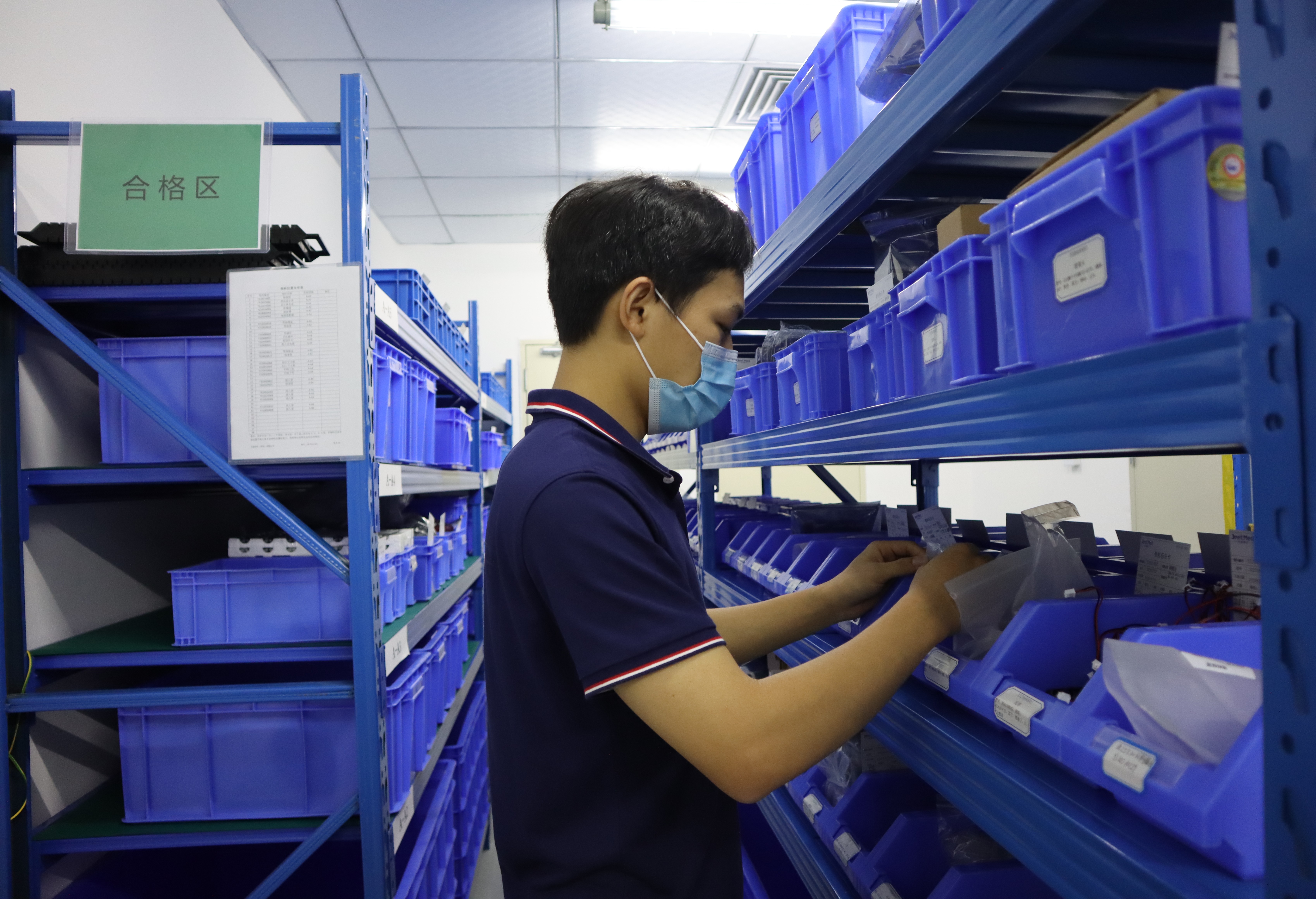
Manufacturing scale behind the specification sheet: flexible veterinary endoscopes are produced and inspected before shipment.
Three practical consequences follow for buyers.
- The accessory ecosystem is growing, and the channel is the gate. As more clinics adopt portable veterinary endoscopes, the range of compatible instruments expands. Whether those instruments can be used depends entirely on the bore size of the scope already owned.
- Two quotes can look identical and behave differently. Outer diameter and resolution are easy to list; channel diameter is the line that decides whether a quoted scope supports the procedures a practice already performs.
- Specification language is becoming standardised. The ISO 8600 series applies to medical endoscopes — ISO 8600-1 for general requirements and ISO 8600-3 for field of view. Buyers writing tenders or comparison sheets increasingly reference this family of standards, and suppliers are expected to respond in the same vocabulary.
What the Working Channel Is and Where It Sits
A flexible veterinary endoscope is built from a small number of functional parts, and the working channel touches almost all of them.
- Control handle. Houses the angulation control. On JeetVet instruments, angulation is driven by a joystick on the handle, and the handle also carries the accessory entry port.
- Insertion tube. The flexible portion that enters the patient. The working channel runs the full length of this tube.
- Bending section. The steerable distal portion that the joystick articulates.
- Distal tip. Contains the camera and light source, and terminates the working channel at an opening beside the optics.
Three relationships follow from this layout, and each has procurement implications.
The channel follows the tube; it does not steer itself. The lumen bends with the insertion tube as the joystick articulates the tip. It cannot be angled independently, and the tube itself must not be bent by hand. That is a handling rule, not a preference: manual bending of the distal end can damage the internal structure.
The channel is a reprocessing surface. Unlike the outer sheath, it is a narrow internal lumen that must be brushed and flushed between patients. The channel diameter determines which cleaning brushes physically pass through it, which is why reprocessing consumables are specified alongside the scope rather than after purchase.
The channel and the optics are independent systems. The RAE-109 gastroscope is documented at 5.8 mm outer diameter with 1,000,000 pixels. That resolution is an optical specification and is unaffected by the scope's 2.0 mm channel. A buyer comparing image quality is comparing optics; a buyer comparing procedural capability is comparing the channel.
What Channel Diameter Actually Controls — Four Clinical Actions
Inside a flexible endoscope, one lumen serves four jobs. That is the core concept behind every channel-size decision.
1. Instrument passage
The channel is a hard physical gate. An accessory that does not fit cannot be used at all, regardless of what the practitioner would prefer. Retrieval instruments and biopsy forceps are limited by bore diameter, and a wider channel widens the range of instruments that can be passed.
2. Flushing and lavage
Fluid is delivered through the channel to clear debris from the field of view or to distend tissue for inspection. A wider bore allows fluid to reach the tip more freely; a narrow bore restricts volume and flow.
3. Suction and aspiration
Fluid, mucus, and debris are drawn back through the same lumen. This is where channel size becomes most visible in daily use: a narrow channel clears slowly and can occlude, particularly with thicker material, while a wider channel recovers the field faster.
4. Air insufflation
Air delivered through the channel opens a collapsed lumen so the clinician can see its walls. It uses the same pathway as flushing and suction, so the three functions cannot run simultaneously — they are sequenced.
The practical consequence is that the channel is a shared resource. A clinician cannot pass a forceps and suction at the same moment. In a busy practice, the speed at which those actions can be sequenced is what determines whether the exam fits into a normal appointment slot.
Step-by-Step: Matching Channel Diameter to Clinical Intent
The following sequence keeps a channel decision anchored to what the instrument must do, rather than to the specification that happens to be listed first.
- Name the anatomy and the species. An ear canal in a small dog and an upper airway in a horse impose very different outer-diameter ceilings. Anatomy sets the hard limit before any other choice is made.
- Decide whether the exam is visual-only or interventional. If the procedure is inspection plus occasional flushing, a small channel is sufficient. If biopsy, retrieval, or foreign-body work is expected, the channel becomes the deciding specification.
- Choose the smallest outer diameter that clears the anatomy. This is the constraint, not the preference. Patient comfort and safe passage come first.
- Within that outer diameter limit, select the largest channel available. Once the anatomy is satisfied, extra bore converts directly into procedural capability — more instruments fit, and flushing and suction are more effective.
- Confirm your actual accessories pass the bore. This step is skipped more often than any other. Procurement teams should list the instruments, forceps, and cleaning brushes the practice already owns and verify that each fits the channel being quoted.
- Confirm reprocessing fits the channel. Check that approved disinfectants and soft-bristled brushes designed for endoscopic equipment are available in the correct size, and confirm the wipe-down rule for the handle.
- Confirm single-operator usability. If one veterinarian must hold the scope, steer it, flush, and suction without an assistant, control design and start-up speed matter as much as bore diameter.
Use Cases: Small Animal, Large Animal, and Field Practice
Small animal otoscopy — RAE-201, 1.2 mm channel
Ear canal examination in dogs and cats is constrained by anatomy: the canal is narrow, and the insertion tube must be slim to pass it safely. The RAE-201 flexible otoscope's 1.2 mm working channel matches that constraint. This configuration supports visual examination with flushing, and it is the right answer when the clinical goal is inspection rather than instrument passage. When the same case moves toward interventional work, the 2.0 mm group is the next step up.
Small animal gastrointestinal work — RAE-109, 2.0 mm channel
The RAE-109 is documented as a gastroscope with a 5.8 mm outer diameter, a 2.0 mm working channel, and 1,000,000 pixels. That combination targets the middle of the range: slim enough for small animal work, with a channel wide enough to support flushing, suction, insufflation, and the passage of instruments designed for a 2.0 mm bore. The RAE-107 and VET-5820 carry the same 2.0 mm channel, which means a practice can standardise on one accessory set across several models in the range.
Large animal and equine work — VET-8528, 2.8 mm channel
The VET-8528 large-animal endoscope carries the widest channel in the portable range at 2.8 mm. Larger patients mean more fluid, more debris, and larger instruments, and a wider bore is what makes those procedures practical. For mobile and ambulatory practice, the same instrument is designed for single-veterinarian operation, so a clinician working alone in the field is not dependent on a second pair of hands.
Portable versus traditional setups
Portability changes how a channel is used. In a fixed clinic, a scope can be set up once and used through the day. In mobile practice, the instrument must be ready quickly at each stop and operated by one person. JeetVet's comparison data documents a 5-second boot-up and single-veterinarian operation, and describes the instruments as suited to clinic, field, and mobile use — three conditions that put a premium on fast start-up and simple tube handling rather than on additional hardware.
Comparison: Working Channel Across the JeetVet Portable Range
| Model | Working channel (inner diameter) | Documented role in the range |
|---|---|---|
| RAE-201 | 1.2 mm | Flexible otoscope |
| RAE-107 | 2.0 mm | Portable flexible endoscope |
| RAE-109 | 2.0 mm | Gastroscope; 5.8 mm outer diameter; 1,000,000 pixels |
| VET-5820 | 2.0 mm | Portable flexible endoscope |
| VET-8528 | 2.8 mm | Large-animal endoscope |
The table shows the practical structure of the range: one narrow-channel model for anatomy-limited examinations, a 2.0 mm group that shares a common accessory set, and one wide-channel model for large-animal procedures. Standardising a clinic or a distribution portfolio on the 2.0 mm group means one set of accessories, one set of cleaning brushes, and one training baseline.
Comparison: Documented Decision Dimensions
Channel diameter is one specification among several that decide whether a purchase works in daily practice. The table below reproduces the comparison dimensions documented in JeetVet's own product comparison data, which positions the portable range against the Karl Storz and Olympus systems named in that documentation.
| Decision dimension | Documented JeetVet position | Benchmark referenced in that data |
|---|---|---|
| Total cost of ownership | 25% lower total cost of ownership | Karl Storz and Olympus comparison set |
| Maintenance cost | 20% lower maintenance cost; low-cost parts | Karl Storz and Olympus comparison set |
| Start-up time | 5-second boot-up | — |
| Operating model | Single-veterinarian operation | — |
| Tube control | Joystick control on the handle; free training provided | — |
| Use environment | All-terrain: clinic, field, mobile | — |
These figures describe JeetVet's own documented positioning relative to the benchmark systems named in that documentation; they are not independent third-party test results, and specifications for the benchmark systems are not reproduced here. Buyers should request current documentation for any system under comparison before finalising a purchase decision.
Read alongside the channel table, the comparison suggests a simple rule for decision-stage buyers: fix the channel size first, because it is the specification that cannot be changed after purchase, then compare cost, maintenance, and service terms within that channel class.
Handling Rules That Protect the Channel
Channel bore is a mechanical feature, and the failure modes that affect it are operational rather than optical. JeetVet's documented handling controls address both.
- Angulation through the joystick only. The insertion tube must be steered exclusively with the joystick on the handle. Manually bending, twisting, or forcing the distal end or the flexible tube is prohibited.
- Stop on resistance. If significant resistance is felt while toggling the joystick, the correct response is to stop immediately to prevent wire breakage.
- Return to neutral before withdrawal. The joystick should be back in the neutral, straight position before the scope is withdrawn, to avoid mucosal trauma and mechanical strain.
- Never immerse the handle. Only the insertion tube is designed for immersion during cleaning. The handle must be disinfected by wiping with dampened alcohol swabs or dedicated wipes, and must not be sprayed directly.
- Store vertically. Vertical storage prevents residual moisture from the tube backflowing into the handle.
- Schedule maintenance. Leak tests and tension wire calibration are specified every 6 months, alongside pre-operation training for staff and neutral-position locking as standard practice.
On the supplier side, the same documentation specifies approved disinfectants, endoscope-specific cleaning solutions, and soft-bristled brushes designed for endoscopic equipment — the consumables that have to match the channel they are cleaning.
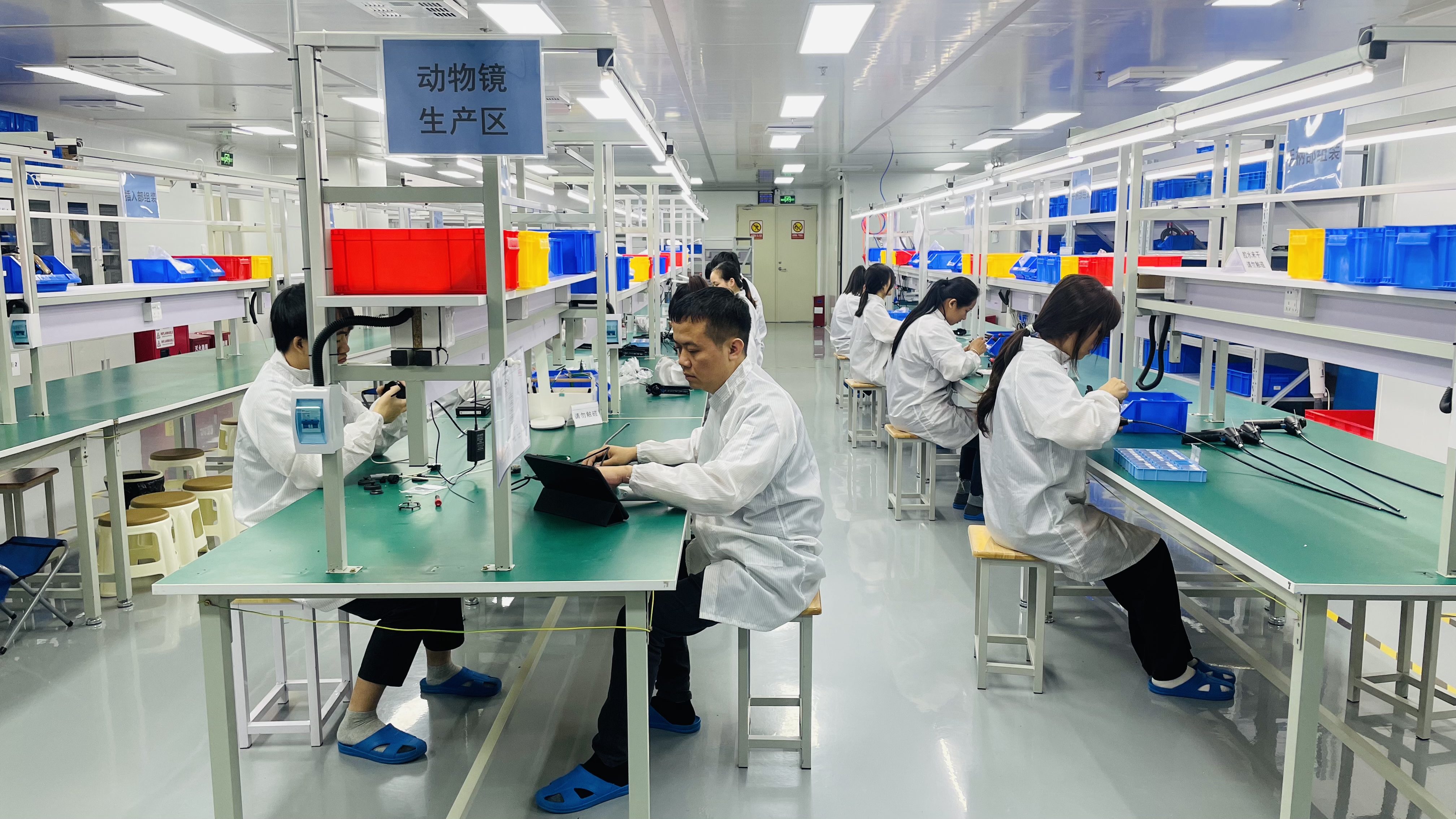
Production and quality control: Jeet Medical operates a 100,000-class cleanroom and a 10,000-class microbiological laboratory.
Documented credentials behind the range include ISO 13485 certification by SGS, a 100,000-class cleanroom, and a 10,000-class microbiological laboratory operated by Jeet Medical (Shenzhen) Co., Ltd. A Computer Software Copyright Registration Certificate (No. 9413508) applies to the Jeet Medical Multi-Camera Automatic Fusion System V1.0. These are quality-system and software credentials; they support the manufacturing claim, and they are separate from the mechanical channel specification discussed in this article.
Frequently Asked Questions
Does working channel diameter change how a veterinary endoscope must be cleaned or certified?
Yes to cleaning, and it depends on the standard. The channel is an internal lumen that must be brushed and flushed, so its diameter determines which cleaning brushes physically pass. JeetVet's documented handling rules separate the two parts of the scope: the insertion tube is designed for immersion during cleaning, while the handle is not waterproof and must be wiped with dampened alcohol swabs or dedicated wipes rather than submerged or sprayed. Approved disinfectants, endoscope-specific cleaning solutions, and soft-bristled brushes are specified. On certification, ISO 13485 (certified by SGS) covers Jeet Medical's quality management system, while the ISO 8600 series — ISO 8600-1 for general requirements and ISO 8600-3 for field of view — is the standards family that applies to medical endoscopes generally.
What can a 2.0 mm working channel do that a 1.2 mm channel cannot?
A wider bore widens the range of accessories that can physically pass and gives more room for flushing fluid, suction, and air to move through the lumen. In the JeetVet range, the 1.2 mm channel belongs to the RAE-201 flexible otoscope, while the 2.0 mm channel is used on the RAE-107, RAE-109, and VET-5820, and the VET-8528 large-animal endoscope carries a 2.8 mm channel. Channel size does not change the optics: the RAE-109 gastroscope is documented at 5.8 mm outer diameter with 1,000,000 pixels regardless of its 2.0 mm channel. The practical rule is to choose the smallest channel that still passes what the procedure requires, because outer diameter and channel bore compete for space inside the same insertion tube.
Does a bigger working channel mean a higher price?
Channel diameter is not the main price driver by itself; optics, portability, and control design generally matter more. Public list pricing is not published for these instruments. What is documented is comparative cost positioning: JeetVet's comparison data states 25% lower total cost of ownership and 20% lower maintenance cost against the Karl Storz and Olympus systems it is benchmarked against, alongside low-cost parts and free training. For a small clinic weighing purchase price against procedural capability, the more useful measure is cost per usable procedure — which is why matching bore to clinical intent matters more than buying the largest channel available.
Can a clinic evaluate a scope before committing to a purchase?
An evaluation should test the three things that only appear in the hand: whether the accessories your clinicians actually use pass the channel, whether one veterinarian can operate the scope unaided, and how the tube responds to the joystick. JeetVet's comparison data documents single-veterinarian operation and a 5-second boot-up, both of which are quick to verify in a hands-on session, and free training is listed alongside low-cost parts. Evaluation units, demonstration arrangements, and their terms are confirmed directly with the supplier rather than published as a fixed policy — the contact details and downloadable brochure at the end of this page are the place to start that conversation.
Does working channel size affect lead time or availability?
Two things drive it: the model configuration and the supplier's production scale. Jeet Medical (Shenzhen) Co., Ltd. is documented as a 5,000 m² facility with 130 employees and an annual output of 10,000 units, exporting 50% of production to Europe, North America, Southeast Asia, Latin America, and the Middle East. Lead time is confirmed per order rather than quoted as a fixed figure, because channel and optical configuration are set at model level — RAE-201, RAE-107, RAE-109, VET-5820, and VET-8528. Practices and distributors standardising multiple sites on the same channel size should confirm scheduling early; the brochure download below and the contact team are the starting point.
Conclusion: A Plain-Language Checklist for Matching Channel to Clinical Intent
The working channel is the specification that cannot be adjusted after purchase. Everything else — resolution, screen, weight, carrying case — can be lived with. Bore size cannot. The checklist below is written for a single decision-maker who has to choose correctly once.
- What will the scope do besides see? If instruments must pass through it, the channel is the deciding number. If the exam is inspection with flushing only, a narrow channel is sufficient.
- How narrow is the anatomy? Set the outer diameter ceiling first, based on species and target site.
- Within that outer diameter, take the largest channel you can get. Once the patient's anatomy is satisfied, extra bore converts directly into capability.
- Confirm your actual accessories fit. List the instruments, forceps, and cleaning brushes in use and check each against the bore before ordering.
- Confirm flushing, suction, and insufflation work at that bore. These three share one lumen, so they compete, and they set the pace of the exam.
- Confirm one veterinarian can run it alone. A 5-second boot-up and joystick tube control matter more in a real appointment slot than they appear to on a datasheet.
- Confirm reprocessing fits. Correct-size brushes, approved disinfectants, wipe-down only for the handle, vertical storage, and 6-month leak testing and tension wire calibration.
- Confirm the supplier's documentation. ISO 13485 certification by SGS, ISO 8600 series reference for specification language, and verifiable production scale.
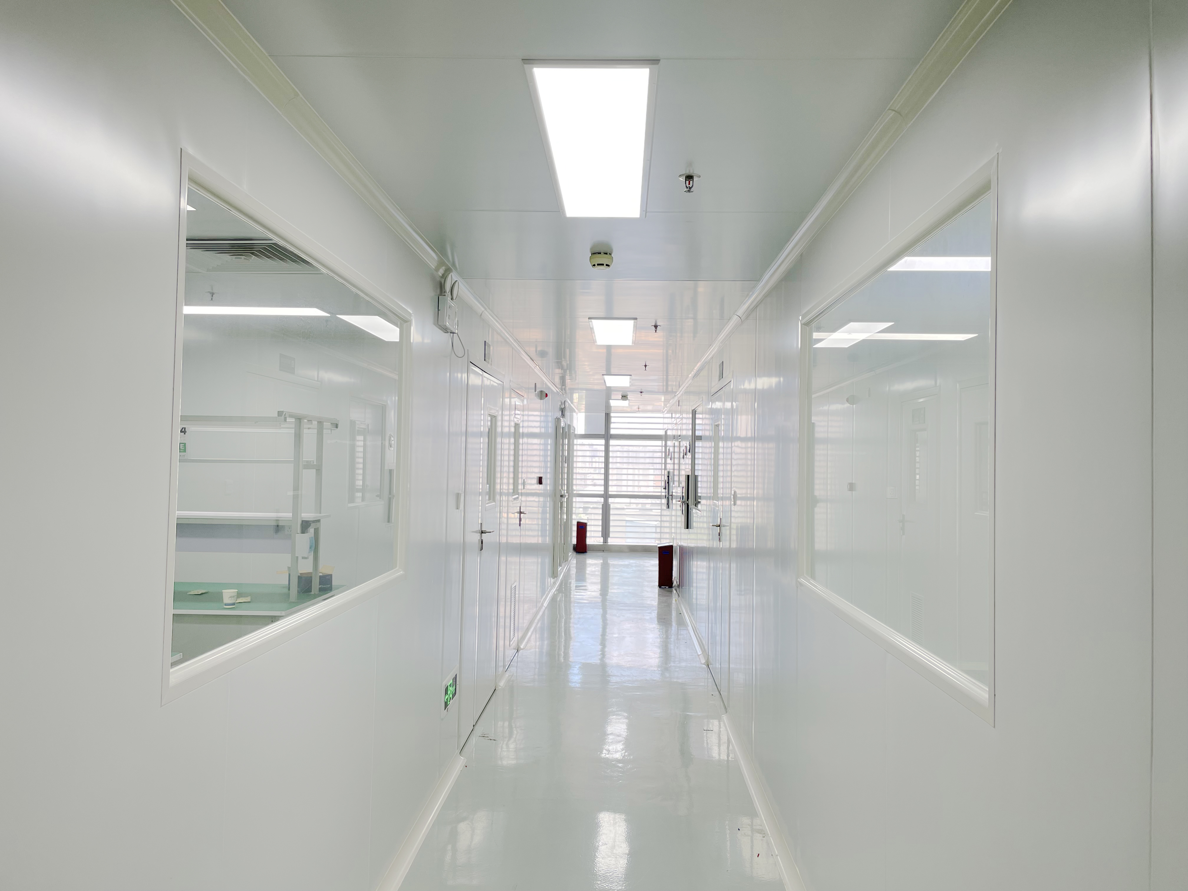
Confirm the channel size against your clinical intent before you order.
Next Step: Check the Channel Against Your Procedures
If you are comparing portable veterinary endoscopes for a clinic, a mobile practice, or a distribution portfolio, start with the channel size and work outward. You can download the full product brochure, review model-level specifications, and confirm which accessories and cleaning consumables match each bore with the JeetVet team.
Brochure download: JeetVet veterinary endoscope product catalog (PDF)
Website: www.jeetvet.com
Email: troy@jeetmed.com / jeetvet66@gmail.com
Telephone: +86 755-23251842 / +86 13381945549
WhatsApp: +86 13816158380