
Cancer Treatment Hospital Evidence: Procedure Records and Imaging Comparisons
Referral evaluators and procurement teams do not assess a cancer treatment hospital the way they assess a device supplier. There is no datasheet. What exists instead is a record trail: dated images, signed procedure notes, pathology results, and a discharge plan that states what happens next. For advanced, recurrent or inoperable disease, that trail is usually the most concrete thing an external evaluator can examine before a treatment date is confirmed.
Guangzhou Fuda Cancer Hospital is an oncology-specialised hospital in Guangzhou, China, operating under the Health Commission of Guangdong Province across two campuses with more than 30,000 m² of floor area, 400 open beds and 45 VIP rooms. It was the first oncology-specialised hospital in Guangdong Province to be accredited by Joint Commission International (JCI). Its relevance to this question is not the length of its therapy menu, but the fact that named treatment steps inside its published cases are attached to dated images and to defined record deliverables.
This article sets out the document set an evaluator can request, shows how that set is tied to named treatment steps in two published cases, and states plainly what those cases cannot prove.
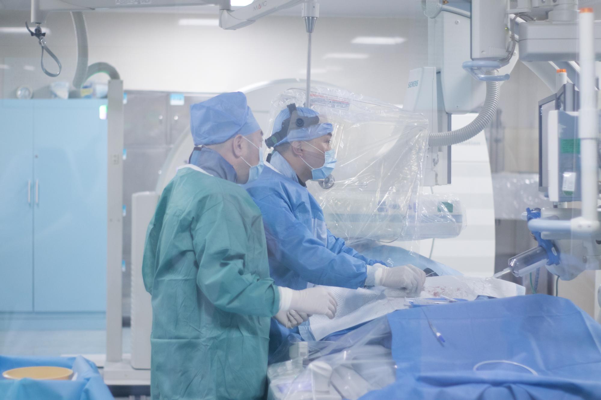
Interventional oncology procedures are performed in an image-guided suite. The equipment and the procedure record are what allow an external evaluator to check that a stated capability corresponds to a dated intervention.
Why procedure records, not treatment menus, decide referral decisions
Cancer treatment capability is easy to list and hard to verify. The same words appear on most provider pages: cryoablation, irreversible electroporation, interventional embolization, immunotherapy. Two hospitals can publish identical therapy lists and still differ substantially in whether those therapies are performed in-house, on which patients, with what imaging guidance, and with what documentation.
The evaluation problem is also predictable. The trigger scenarios are usually imaging-detected progression or recurrence, or the failure of a prior treatment line. Urgency is often high, because patients with progressive or symptomatic tumors need timely evaluation and intervention, and some cases are time-sensitive.
The underlying clinical constraints repeat themselves as well. Tumors are frequently locally unresectable, disease may already be systemic, and there is a need to reduce systemic toxicity or to preserve organ function with less invasive options. Common challenges include tumors adjacent to critical structures that prevent surgery. Those constraints are exactly why documentation matters: when a conventional pathway is closed, the next option has to be justified by something an evaluator can read.
The record set a hospital can be asked to produce
Deliverables are the unit of verification. In specialised oncology care, the documented outputs of an episode of care are a personalized treatment plan, imaging and pathology reports, treatment records with a follow-up schedule, and rehabilitation and nursing recommendations.
For the interventional cases published by Fuda, the deliverable set is stated more narrowly and is therefore easier to check: imaging comparison reports, procedure records, a discharge and follow-up plan, and nursing records. These can be requested from the care process of a specific case.
| Document | What it records | What it does not establish on its own | How an evaluator verifies it |
|---|---|---|---|
| Imaging comparison report | Pre-treatment and post-treatment imaging for a named lesion, with dates | That the studies belong to the named patient, unless identifiers are cross-checked | Compare image dates against the procedure record; request source imaging |
| Procedure record | Technique used, guidance modality, anesthesia approach, session structure | Clinical appropriateness of the same technique for a different patient | Match technique and procedure date to the treatment plan |
| Pathology / biopsy result | Tissue diagnosis and, where performed, molecular markers | Response to treatment | Cross-check with the MDT note and the imaging timeline |
| MDT note / treatment plan | Which disciplines reviewed the case and what was planned, in what order | That the plan was executed as written | Compare plan against procedure and discharge records |
| Discharge and follow-up plan | Post-discharge instructions, review intervals, escalation triggers | Long-term outcome | Confirm that later follow-up records exist |
| Nursing records | Inpatient monitoring, symptom management, complications | Procedure technique | Read alongside the procedure record for the same dates |
How the documented service pattern is structured at Fuda
Fuda's clinical model is described as the 3C+P model: Cryo-Irreversible Electroporation Ablation (CIA), Cancer Vascular Intervention (CVI), and Combined Immunotherapy for Cancer (CIC), plus Personalized (P) comprehensive therapy. Multidisciplinary team (MDT) planning runs across the model rather than sitting inside a single department, which is why the MDT note appears in the record set rather than as background paperwork.
The equipment base named for these procedures is 64-slice CT, 1.5T MRI and DSA, together with NanoKnife and cryoablation devices. Irreversible electroporation (NanoKnife) was approved for clinical application in China in June 2015, and Fuda Cancer Hospital was the first to introduce the therapy. The stated service scope runs from diagnostics — imaging, tumor markers, biopsy and genetic testing — through local therapies including cryoablation, IRE, microwave ablation and radiofrequency ablation, to interventional treatments such as HAIC, TACE and TAI with drug-eluting microspheres, immuno- and cell therapies, rehabilitation, psychosocial support and follow-up.
For cross-border cases, the hospital operates international patient care support with service in English, Thai, Indonesian, Malay, Russian, Kazakh, Mongolian, Chinese and Cantonese. Verified third-party data records more than 10,000 international cancer patients from 100+ countries, with 60% of patients drawn from Southeast Asia, the Middle East, Europe and North America. Language coverage and record assessment are part of the evidence chain, not a separate hospitality layer: an evaluator who cannot read a record cannot verify it.
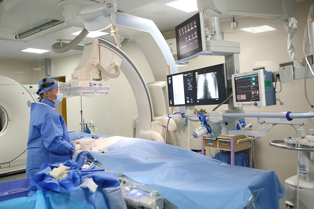
DSA and multidetector CT systems support both the intervention and the imaging comparison that follows it. The imaging report is one of the four documented deliverables an evaluator can request.
Case file 1: single-needle, local-anesthesia nanoknife ablation in recurrent pancreatic cancer
The clearest example of records being tied to named steps is a pancreatic case published as ‘Single-needle local anesthesia pancreatic nanoknife ablation’. The patient was an 81-year-old man from Hong Kong, China, with recurrent pancreatic cancer after a Whipple procedure. Imaging identified an approximately 7 cm irregular hypoechoic mass in the retroperitoneum encasing the celiac trunk and the superior mesenteric artery, and suggested non-resectability. Advanced age and poor cardiopulmonary reserve made resection or general anesthesia intolerable for this patient.
The procedure performed was percutaneous single-needle bipolar nanoknife (S-IRE) ablation under CT and ultrasound guidance, carried out under local anesthesia, with concurrent biopsy and celiac plexus block. In practical terms, ablation, pathology sampling and pain control were combined in a single session rather than distributed across separate appointments — a pattern that shows up directly in how the procedure is documented.
The execution sequence is written down step by step: pre-operative MDT, image-guided puncture, single-needle energy delivery, concurrent biopsy and celiac plexus block, then post-operative imaging review and short rehabilitation. The published deliverables for the episode are an intra- and post-operative imaging comparison, pathology results, recovery records and follow-up recommendations.
The content of the imaging comparison is specific rather than generic. Pre-treatment CT imaging dated August 5 and post-treatment CT imaging dated August 14 are attached to the official case report, together with procedural photographs. The reported patient outcome was ambulation the next day and CT evidence of necrosis and shrinkage of the lesion. No numeric measurement of that shrinkage was published; the case reports the finding qualitatively, and an evaluator should record it that way.
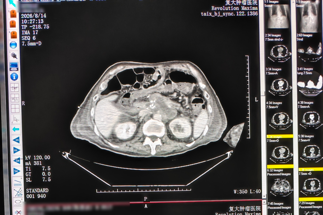
Post-treatment CT imaging dated 14 August, published as part of an anonymized case report on single-needle nanoknife ablation. It forms one half of a pre/post comparison pair whose pre-treatment study is dated August 5.
Case file 2: MDT-planned treatment for a 14.4 cm liver lesion
The second case is about planning logic rather than a single procedure. Published as ‘Regular checkups yet late-stage liver cancer found: why was a 14.4cm tumor missed?’, it involved a vehicle mechanic from Guangxi, China, who traveled to Guangzhou for care. Diagnosis identified primary liver cancer with multiple intrahepatic metastases, the largest lesion measuring approximately 14.4 cm with areas of liquefactive necrosis, and markedly elevated AFP. Routine checkups had not detected the disease, and local care options were limited because the patient had a contrast allergy.
The documented solution had a sequence: contrast desensitization before image-guided hepatic arterial infusion chemotherapy (HAIC), HAIC combined with targeted therapy and immunotherapy, and later evaluation for possible conversion to ablation or surgery. Two HAIC sessions were completed shortly after admission, with further therapy and checks scheduled according to imaging results and the patient's condition.
Reported results were a tumor reduction of approximately 5 cm after two treatments, disappearance of the original tumor thrombus, and a decline in AFP to the normal range as reported in the case. The documented deliverables for this episode are comparative imaging — admission versus post-two-HAIC — MDT notes, and treatment and follow-up records.
This is the case that shows why an MDT note is an evidence document rather than a formality. The desensitization step, the choice of an intra-arterial route, and the stated intention to reassess for conversion to ablation or surgery only make sense when the reasoning is recorded. An evaluator reading the file can see which decision was made, when, and on what basis.
| Published case | Named treatment steps | Evidence attached to the report | Records that can be requested | Boundary stated in the case |
|---|---|---|---|---|
| Single-needle local anesthesia pancreatic nanoknife ablation | Pre-operative MDT → image-guided puncture → single-needle energy delivery → concurrent biopsy and celiac plexus block → post-operative imaging review and short rehabilitation | Pre-treatment CT dated August 5, post-treatment CT dated August 14, procedural photographs | Intra- and post-operative imaging comparison, pathology results, recovery records, follow-up recommendations | No numeric values were published for lesion shrinkage |
| Regular checkups yet late-stage liver cancer found (14.4 cm lesion) | Admission assessment → MDT → desensitization and HAIC planning → two HAIC sessions → imaging assessment and AFP monitoring → continued targeted immunotherapy with conversion assessment | Comparative imaging (admission versus post-two-HAIC), MDT notes | Treatment and follow-up records, MDT notes, comparative imaging | Results derive from a single case; conversion to ablation or surgery was still under evaluation |
What this evidence does not establish
Published case reports are anonymized before release, and every claim in this article is matched to the case report rather than extended beyond it. An evaluator should hold the same line. Four limits are worth stating explicitly.
Publication is not the medical record. Published cases identify findings, dates and outcomes, but original imaging and pathology reports require provision or authorization by the hospital for medical proof. A case page is not a radiology report, and an evaluator who needs proof should request the source file.
A single case is not a population. A lesion shrinking from nearly 17 cm to about 5 cm in a published Ewing sarcoma lung metastasis case, or a near-50% radiologic reduction within about two months in a metastatic breast cancer case, describes those individual patients. Outcomes vary between patients, and case-selection effects are real.
Some published values still require verification. In a published lung cancer case involving a Malaysian patient, the record states that imaging showed lesion shrinkage and that the tumor marker CYFRA21-1 decreased to normal, with the explicit note that the exact values require verification in the medical records.
Applicability is constrained by anatomy and by prior treatment. The local-anesthesia, single-needle approach in the pancreatic case was selected because imaging suggested non-resectability and because the patient could not tolerate resection or general anesthesia; it is documented as an individual case, not as a general indication for pancreatic lesions. More broadly, conventional methods have restricted applicability for unresectable tumors or tumors adjacent to critical structures, and ablation addresses local disease. Systemic metastases still require systemic therapy, which is why the liver case combined HAIC with targeted therapy and immunotherapy instead of relying on a local technique alone.
Market signals behind the demand for documentation
The demand for verifiable records is not only a clinical preference. It tracks the size and direction of the market.
China's hospital services market was estimated to reach USD 614.82 billion in 2024, with specialized private hospitals representing a significant growth driver on the back of an aging population and rising healthcare expenditure. Cancer treatment accounts for a visible share of that spend: China's healthcare expenditure for cancer treatment reached 221.4 billion RMB, or 5.4% of total health expenditure.
On the technology side, the global cryoablation devices market was projected to grow from USD 614.3 million in 2026 to USD 1,036.7 million by 2030, with hospitals as the largest end-user segment at 54.4%. Clinical evidence accumulates in the same direction: a Fuda Cancer Hospital study cited by an international expert consensus reported a 100% technical success rate for complete ablation of subsolid nodules across 19 cases.
Read together, these figures describe a market in which ablation and interventional devices are deployed inside hospitals rather than simply sold to them. That shifts the differentiating asset toward the hospital's own procedural record. A buyer evaluating an interventional oncology provider is therefore evaluating documentation quality as much as equipment inventory.
Future outlook
Two shifts are reasonable to expect. The first is standardisation of the evidence pack: imaging comparison reports, procedure records, discharge and follow-up plans, and nursing records requested as a defined set rather than assembled case by case. The second is remote review. Fuda already offers initial remote consultations and assessments through online appointment and tele-evaluation, alongside its international patient service center, which supports remote medical record assessment.
For referral partners, that changes the practical question from ‘what does this hospital offer?’ to ‘what will this hospital show me, in what format, and how quickly?’
A practical checklist for evaluating a hospital evidence pack
- Request the imaging comparison report first, and confirm that both the pre-treatment and post-treatment studies carry dates.
- Match the procedure record to the treatment plan: same date, same technique, same guidance modality.
- Establish whether ablation, biopsy and pain control were performed in one session or across separate sessions, and whether the record states this explicitly.
- Read the MDT note for the reasoning behind the sequence, not only for its conclusion.
- Check the discharge and follow-up plan for review intervals and escalation triggers.
- Request the pathology or molecular result on which any systemic therapy choice was based.
- Ask what the published case does not state, including whether numeric outcome values were released.
- Confirm how original imaging and pathology reports can be obtained and authorised.
- For international cases, confirm language coverage and whether record assessment can be completed before travel.
- Treat a therapy name without a dated record as unverified.
FAQ
What is the difference between a hospital's therapy list and its procedure records?
A therapy list names techniques the hospital states it offers. A procedure record documents what was performed for a specific patient on a specific date, including the guidance modality, the anesthesia approach and the session structure. In interventional oncology, the documented deliverable set is imaging comparison reports, procedure records, a discharge and follow-up plan, and nursing records. Only the second category can be cross-checked against images and dates.
Which documents can a referral evaluator request before confirming a treatment date?
The stated deliverables of an episode of care are a personalized treatment plan, imaging and pathology reports, treatment records with a follow-up schedule, and rehabilitation and nursing recommendations. In published interventional cases the narrower set is imaging comparison reports, procedure records, a discharge and follow-up plan, and nursing records, which can be requested from the care process of a specific case.
What does an imaging comparison report in an interventional case contain?
It pairs pre-treatment and post-treatment imaging for a named lesion, with dates. In the published pancreatic case, pre-treatment CT imaging dated August 5 and post-treatment CT imaging dated August 14 were attached to the official case report alongside procedural photographs. The comparison supports a description of change, such as evident necrosis and shrinkage of the lesion. It does not by itself establish patient identity, and it does not establish that the technique is appropriate for a different patient.
How is a session that combines ablation, pathology sampling and pain control documented?
As a single execution sequence. In the pancreatic case the documented steps were pre-operative MDT, image-guided puncture, single-needle energy delivery, concurrent biopsy and celiac plexus block, then post-operative imaging review and short rehabilitation. The published deliverables were an intra- and post-operative imaging comparison, pathology results, recovery records and follow-up recommendations. Because the plexus block and the biopsy occur in the same session as the ablation, the record has to show all three steps rather than the ablation alone.
Why are published cancer case reports anonymized, and what can they not prove?
Cases are anonymized before release to protect patient identity. As a result, published images and narrative cannot substitute for the medical record: original imaging and pathology reports require provision or authorization by the hospital. A published case also cannot demonstrate that an outcome generalizes, because it describes one patient, and some reported values may still need verification in the medical records, as stated in the CYFRA21-1 example.
What should an international patient's record package include at discharge?
For the interventional cases described, the discharge and follow-up plan sits alongside imaging comparison reports, procedure records and nursing records. The wider service deliverable set adds a personalized treatment plan, imaging and pathology reports, a treatment records and follow-up schedule, and rehabilitation and nursing recommendations. For cross-border cases, record assessment can also be requested remotely through the international patient service center before travel.
Reference material: the hospital's English-language brochure is available for download — Fuda Cancer Hospital brochure (PDF).