
Comparing Four Minimally Invasive Cancer Case Profiles
Four published, anonymized case reports describe four different advanced or recurrent cancers treated through non-surgical, image-guided routes at one oncology-specialised hospital in Guangzhou. Read side by side as evidence rather than promotion, they show what a documented minimally invasive cancer treatment record actually contains — and exactly where those records stop.
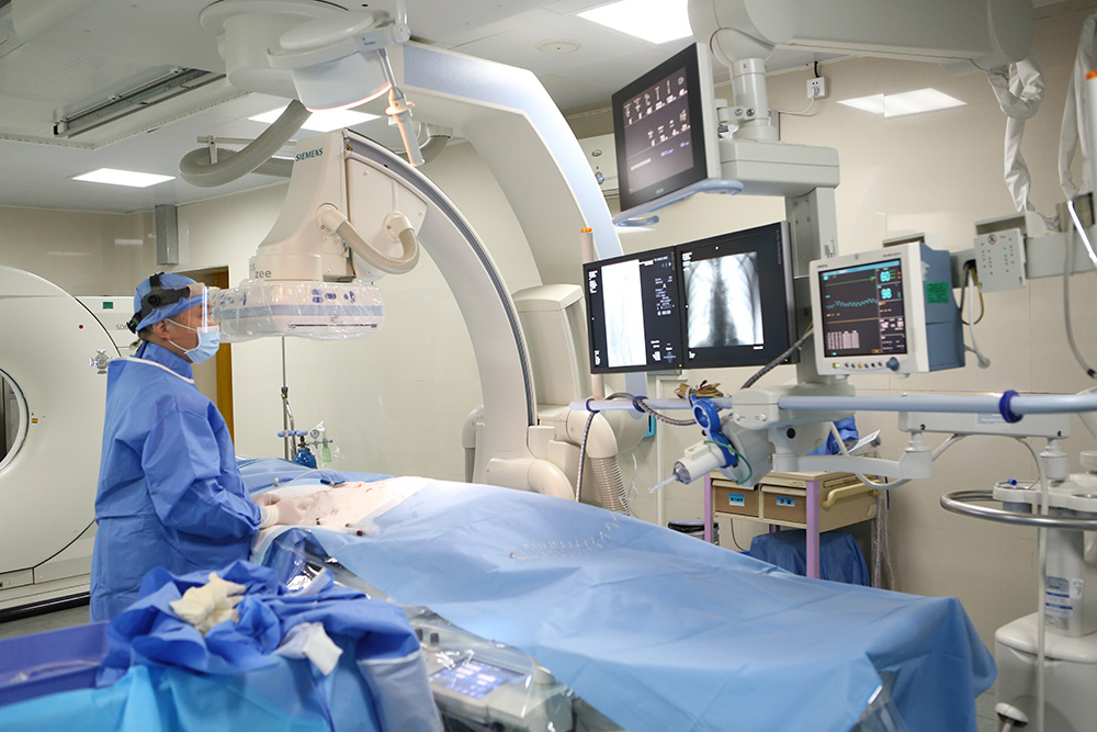
Image-guided oncology workflow: DSA and multi-detector CT imaging support the planning and guidance of minimally invasive, non-surgical tumour treatment.
Minimally invasive cancer treatment is usually discussed in the language of technology: which device, which energy source, which access route. Buyers of oncology care work with a different problem. A referring physician, a hospital partner, an insurer or an international patient in the evaluation stage cannot test-drive a treatment. The closest equivalent to a specification sheet in this field is a published case record — imaging, pathology, a treatment log and a follow-up plan — and the value of that record depends on how completely it was written.
Guangzhou Fuda Cancer Hospital is an oncology-specialised hospital in Guangzhou, China, operating the Tianhe Campus and Haizhu Campus with more than 30,000 m² of floor area, 400 open beds and 45 VIP rooms. It holds Joint Commission International (JCI) accreditation, passed in 2014 with a later re-evaluation mentioned in its documentation, and National Clinical Key Specialty designation in oncology, documented in National Health Commission notice Guo Wei Ban Yi Han [2018] No. 292, alongside Guangdong Provincial Key Clinical Oncology Specialty designation, documented with a certificate dated December 2011.
Four of the hospital's anonymized case reports cover recurrent pancreatic cancer, Ewing sarcoma with lung metastasis, late-stage liver cancer and metastatic triple-negative breast cancer. This analysis compares them on the dimensions a buyer can actually verify: diagnosis complexity, treatment route, documented outcome and available proof assets.
One caution applies before the comparison begins. The four reports use different measurement methods, different follow-up windows and different tumour types. No ranking is offered here, because the underlying documents do not support one: a diameter reduction in one case cannot be ranked against an AFP normalisation in another.
Why Case Profiles Are the Right Evidence Unit at the Evaluation Stage
A case report answers a narrow question — what was done in this specific patient, and what was observed afterwards. It does not answer what usually happens across a population. That distinction matters for buyers, because in minimally invasive oncology the interesting variation is rarely the device; it is the decision sequence.
Four fields determine how much a case report is worth to an evaluator. First, diagnosis complexity: how difficult was the presentation, and what made standard management difficult. Second, treatment route: the actual sequence of interventions, including the preparatory steps that are easy to omit from promotional summaries. Third, documented outcome: whether the reported change is a measured value, an imaging description, or a qualitative statement. Fourth, proof assets: whether the reader can request the underlying imaging and pathology, or whether the claim ends at the article.
Judged on those four fields, the four Fuda case reports are not interchangeable. They differ in how completely each one closes the evidence loop.
The Common Structure Across All Four Documented Cases
Before the differences, the shared sequence is worth stating, because it is the part that transfers between cases. All four records describe the same general architecture, published in the hospital's case documentation:
- A multidisciplinary team (MDT) assessment precedes treatment, rather than a single-specialty decision.
- Local tumour control is delivered through image-guided, non-surgical procedures.
- Systemic therapy — targeted, immunotherapy, ADC-based or chemotherapy — is combined with the local treatment rather than used alone.
- Response is reassessed with imaging, and in one case with tumour-marker monitoring, before the next step is planned.
- Cross-border logistics are documented for the international patients, including admission evaluation, procedure records and discharge planning.
This structure is the reason the four cases can be compared at all. What changes between them is the combination, and that is a function of the tumour, the anatomy and the patient's physiological reserve.
Case Profile 1 — Recurrent Pancreatic Cancer, 81-Year-Old Patient From Hong Kong
Diagnosis complexity. The patient was an 81-year-old man from Hong Kong, China, who received treatment in mainland China. He had previously undergone a Whipple procedure and presented with recurrence in the surgical bed: an approximately 7 cm irregular hypoechoic retroperitoneal mass encasing the celiac trunk and the superior mesenteric artery. Imaging suggested non-resectability, and the report states that advanced age and poor cardiopulmonary reserve made resection or general anaesthesia intolerable. Two constraints applied at once — an anatomically hostile target and a patient who could not tolerate a major procedure.
Treatment route. The documented approach was percutaneous single-needle bipolar NanoKnife (S-IRE) ablation under combined CT and ultrasound guidance, performed under local anaesthesia, with concurrent biopsy and a celiac plexus block. Preoperative MDT review preceded the procedure, and real-time dual-image guidance was used during energy delivery. The anaesthetic choice was therefore not a preference but a consequence of the cardiopulmonary assessment.
Documented outcome. The report states that the patient ambulated the following day and that post-procedure CT showed evident necrosis and shrinkage of the lesion. No numeric tumour measurement is given — a point that matters directly when this case is placed beside the others. Qualitative findings recorded were good recovery, satisfactory pain control and positive family feedback.
Proof assets. The published report includes pre-treatment and post-treatment CT images, ablation process images, pathology results, recovery records and follow-up recommendations, with a photograph of the patient with the lead surgeon.
Case Profile 2 — Ewing Sarcoma With Lung Metastasis, Patient From Lebanon
Diagnosis complexity. The case involved an international patient travelling from Lebanon to Guangzhou for treatment as an individual case. Diagnosis confirmed Ewing sarcoma lung metastasis, and the report notes that pathological differentiation here is complex and can be confused with small cell lung cancer morphology. The pulmonary lesion grew from 8 cm to approximately 17 cm at its largest dimension and sat adjacent to the pericardium and major vessels — an inoperable position that required a non-surgical local approach.
Treatment route. The documented solution was superselective interventional embolization-chemotherapy, followed by combined targeted therapy with periodic CT follow-up. The methodology combined MDT assessment with image-guided superselective embolization delivering local high-concentration chemotherapy, delivered across multiple staged sessions rather than a single procedure.
Documented outcome. The pulmonary metastatic lesion reduced from approximately 17 cm to approximately 5 cm over multiple staged interventional treatments. Tolerance was reported as good: symptom relief, no significant vomiting and only mild skin dryness. Patient feedback quoted in the case was that the patient had made the right choice.
Proof assets. The case article includes CT images and treatment comparison figures, supported by imaging comparison reports, procedure records, a discharge and follow-up plan, and nursing records.
Case Profile 3 — Late-Stage Liver Cancer, Patient From Guangxi
Diagnosis complexity. The patient was an individual from Guangxi who travelled to Guangzhou for treatment. He worked as a vehicle mechanic and was the family breadwinner from a low-income background. Diagnosis identified primary liver cancer with multiple intrahepatic metastases, a largest tumour measuring approximately 14.4 cm with areas of liquefactive necrosis, and a markedly elevated AFP level that was later reported to decrease. Two complications shaped the case: the cancer was detected despite routine checkups, and local care options were limited because the patient had a contrast allergy — a common constraint on image-guided vascular procedures.
Treatment route. A contrast desensitization step was performed before image-guided hepatic arterial infusion chemotherapy (HAIC). Two HAIC sessions were completed shortly after admission, combined with targeted and immunotherapy, followed by imaging assessment and AFP monitoring. The plan then continued with targeted immunotherapy and an assessment of whether the tumour could be converted to ablation or surgery.
Documented outcome. The tumour reduced by approximately 5 cm after two treatments, the original tumour thrombus disappeared, and AFP declined to the normal range as reported in the case. Symptom relief and improved quality of life were recorded, and the family received assistance through a hospital charity fund.
Proof assets. Comparative imaging (admission versus after two HAIC sessions), MDT notes and treatment and follow-up records are referenced, together with a letter of thanks. The report states that original images and medical records can be requested from the hospital for verification.
Case Profile 4 — Metastatic Triple-Negative Breast Cancer, Patient From Indonesia
Diagnosis complexity. The patient was an international patient from Indonesia who travelled to Guangzhou for care. Diagnosis identified metastatic triple-negative breast cancer with lesions in bone, pleura, subcutaneous tissue, liver and meninges, and in-hospital pathology and molecular testing noted low HER2 expression. Prior multi-line chemotherapy and radiotherapy had been ineffective, and the patient presented with multiple systemic metastases, pulmonary artery thrombosis and severe functional decline — a combination that narrows both surgical and single-modality options.
Treatment route. The documented sequence prioritized thrombus and infection management first, then biopsy and cryoablation of the chest wall lesion, then Gamma Knife treatment for meningeal metastasis, then ADC combined immuno/targeted therapy based on the low HER2 expression identified during in-hospital testing. The methodology was an MDT individualized plan integrating image-guided ablation, stereotactic radiotherapy and molecular marker-guided systemic therapy. Local interventions plus two systemic treatment cycles with imaging reassessment were completed in about two months.
Documented outcome. Overall tumour lesions showed approximately near-50% radiologic reduction within about two months, described as 'nearly halved' in the case article. Pain, dyspnoea and exercise tolerance improved significantly, with the patient progressing from wheelchair dependence to walking longer distances. The family expressed gratitude for the clinical team's professionalism and care.
Proof assets. The case page includes pre-treatment and post-treatment imaging comparisons. The report notes that original imaging and pathology reports need to be provided by the hospital for medical proof.
Side-by-Side Comparison of the Four Documented Cases
| Case | Diagnosis complexity | Documented treatment route | Documented outcome (as reported) | Proof asset status |
|---|---|---|---|---|
| Recurrent pancreatic cancer (81-year-old, Hong Kong) | ~7 cm retroperitoneal recurrence encasing celiac trunk and superior mesenteric artery; imaging suggested non-resectability; age and cardiopulmonary reserve precluded general anaesthesia | Preoperative MDT; CT+US-guided percutaneous single-needle bipolar NanoKnife (S-IRE) ablation under local anaesthesia; concurrent biopsy and celiac plexus block | Ambulatory the next day; CT showed evident necrosis and shrinkage; no numeric measurement given | Pre/post CT images, pathology, recovery records and follow-up recommendations in the published report |
| Ewing sarcoma lung metastasis (patient from Lebanon) | Lesion grew from 8 cm to ~17 cm, adjacent to pericardium and major vessels; pathological differentiation can be confused with small cell lung cancer morphology | MDT assessment; superselective interventional embolization-chemotherapy, then combined targeted therapy with periodic CT follow-up | ~17 cm to ~5 cm over multiple staged treatments; good tolerance, no significant vomiting, mild skin dryness | CT images and treatment comparison figures; procedure records, discharge and follow-up plan |
| Late-stage liver cancer (patient from Guangxi) | ~14.4 cm primary tumour with intrahepatic metastases and liquefactive necrosis; contrast allergy limited local care; AFP markedly elevated | Desensitization then HAIC (two sessions) with targeted and immunotherapy; continued treatment with assessment for conversion to ablation or surgery | ~5 cm tumour reduction; original tumour thrombus disappeared; AFP declined to normal range | Comparative imaging, MDT notes, treatment and follow-up records; originals available on request |
| Metastatic triple-negative breast cancer (patient from Indonesia) | Bone, pleural, subcutaneous, liver and meningeal metastases; prior multi-line chemo/radiotherapy ineffective; pulmonary artery thrombosis | Thrombus and infection management; cryoablation of chest wall lesion; Gamma Knife for meningeal metastasis; ADC combined immuno/targeted therapy | Near-50% radiologic reduction over about two months; improved pain, dyspnoea and exercise tolerance | Pre/post imaging comparisons; original imaging and pathology require hospital provision |
Technical Explanation — Why the Four Treatment Routes Differ
The four routes diverge because each modality has a different relationship to the tumour. Cryoablation destroys tissue through rapid freeze-thaw cycles delivered by probes placed into the lesion; it is suited to accessible solid deposits such as the chest wall lesion in the breast cancer case. Irreversible electroporation, delivered in this hospital's pancreatic case through a single-needle bipolar NanoKnife system, uses electric fields rather than heat or cold, which is relevant when a lesion sits against major vessels — as it did with the celiac trunk and superior mesenteric artery. Vascular interventional oncology, including superselective embolization and HAIC, works through the tumour's blood supply and was used in both the Ewing sarcoma and liver cases. Gamma Knife radiosurgery delivers focused radiation and addressed meningeal metastasis in the breast cancer case, where a lesion was not reachable by needle.
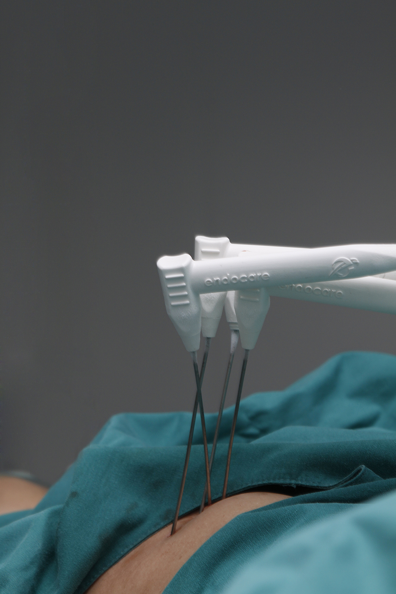
Cryoablation process documentation: freeze-thaw ablation is one of several image-guided local techniques used in non-surgical tumour treatment.
Regulatory context supports the maturity of some of these routes rather than the novelty. Cryosurgery for cancer treatment was approved by China's SDA in 1999, according to hospital documentation. Hospital-published information states that more than 10,000 cryosurgery cases across 30 or more cancer types have been completed at Guangzhou Fuda Cancer Hospital, and the institution's research on cryoablation for lung nodules was cited in the 2024 AATS Expert Consensus. Those are institution-sourced and consensus-level recognitions rather than independent outcome audits.
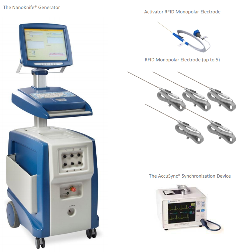
Irreversible electroporation equipment: the NanoKnife approach uses electric fields rather than thermal energy, which matters for lesions adjacent to major vessels.
The practical implication for buyers is that the device list is the least informative part of a capability statement. The hospital's own service documentation describes a technology stack combining 64-slice CT, 1.5T MRI, DSA, cryo devices and NanoKnife, supported by dedicated interventional oncology and ablation teams. What the four cases add is the decision layer above the equipment: which modality was chosen, in what order, and why the sequence changed from patient to patient.
How Minimally Invasive Routes Compare With Surgery-First Management — and Where the Boundary Sits
Surgical resection remains the reference standard where it is feasible and where the patient can tolerate it. The four documented cases are not a substitute argument for that. In the pancreatic case, the lesion encased the celiac trunk and superior mesenteric artery and imaging suggested non-resectability. In the Ewing sarcoma case, the lung lesion sat adjacent to the pericardium and major vessels in an inoperable position. In the breast cancer case, prior multi-line chemotherapy and radiotherapy had failed and the disease was systemically distributed. In the liver case, the tumour burden was large and multifocal. Each case entered minimally invasive treatment because the conventional route was closed or insufficient on its own.
Two boundaries follow from this and should be stated plainly. First, local ablation treats local disease; none of the four cases relied on ablation alone, and all four combined local treatment with systemic therapy. Second, the reported outcomes are individual results, not comparative performance. These reports contain no head-to-head comparison against surgery, no control group and no survival statistics, so they cannot be used to argue that a minimally invasive route outperforms surgery in any of these four tumour types.
Where the minimally invasive approach does show a documented advantage is in specific constrained situations: an anatomically difficult target, an anaesthetic contraindication, or a complication such as the contrast allergy in the liver case or the pulmonary artery thrombosis in the breast case, both of which had to be managed before tumour-directed treatment could proceed at all.
Market Context
The direction of the market is documented, even if outcome-level benchmarking remains thin. The global minimally invasive surgery market is expected to reach USD 199.30 billion by 2030, based on a forecast period of 2025 to 2030 published by MarketsandMarkets. The wider global cancer therapy market is projected to reach USD 700.09 billion by 2034, according to DelveInsight. Regionally, Asia Pacific dominated the cancer treatment facilities market with a 37.6% revenue share in 2025, as reported by Grand View Research.
That last figure is consistent with the geography visible in the four case reports: two domestic patients from Hong Kong and Guangxi, and two international patients from Lebanon and Indonesia. The hospital states that patients from more than 130 countries and regions have sought care there, with 60% originating from Southeast Asia, the Middle East, Europe and North America. The available public data does not extend to service revenue or outcome benchmarking for individual providers, which is precisely the gap that makes case-level evidence the practical unit of evaluation for buyers.
What the Four Reports Do Not Establish
- No comparative or population-level conclusions. Four single-patient reports with no control group cannot establish relative effectiveness against other treatment options.
- Non-standardized outcome metrics. One case reports a diameter change, one reports AFP normalisation, one reports an approximate percentage reduction described as 'nearly halved', and one reports evident necrosis and shrinkage with no numeric value at all. These are not interchangeable measurements.
- Unequal follow-up windows. The breast cancer case describes reassessment at about two months; the pancreatic case reports short postoperative observation; the Ewing sarcoma case describes multiple staged treatments with longer follow-up. Long-term durability is not documented in the published material reviewed here.
- Publication selection. Case reports are chosen for publication by the institution. Cases with unremarkable or unfavourable trajectories are not represented, and no denominator is provided against which these four can be positioned.
- Verification requires authorization. Imaging and pathology originals exist, but in more than one case they must be requested from the hospital, which normally requires patient consent.
- Eligibility is tumour-specific. Ablation and embolization require a targetable lesion and a workable access route. None of the four reports supports a general claim that minimally invasive treatment is suitable for every patient with inoperable or metastatic disease.
Future Outlook
The most likely development in this field is not a new energy source but better documentation. Buyers evaluating oncology providers increasingly ask for the same fields across every case: tumour type and stage, prior treatment, the reason the conventional route was closed, the intervention sequence, a measured outcome at a defined time point, and a retrievable proof asset. Two of the four cases reviewed here partially meet that standard and two fall short on measured outcomes.
A second trend concerns the combination layer. Each of the four cases succeeded or progressed because a local procedure and a systemic regimen were sequenced rather than used in isolation — cryoablation plus Gamma Knife plus ADC therapy in one case, HAIC plus targeted immunotherapy in another, embolization-chemotherapy plus targeted therapy in a third. As molecular testing becomes routine, the selection of the systemic component is likely to become more discriminating, while the local component remains determined by anatomy. That split is where comparative evidence would be most valuable and where it is currently thinnest.
FAQ
What do the four documented case profiles have in common?
All four began with multidisciplinary team assessment, used image-guided local treatment rather than open surgery, combined that local treatment with systemic therapy, and reassessed response with imaging before planning the next step. Two of the four patients also required preparatory management of a complication — contrast desensitization in the liver cancer case and thrombus and infection control in the breast cancer case — before tumour-directed treatment could begin.
Which of the four cases reported the largest measurable change?
The reports are not directly comparable because they measure different things. The Ewing sarcoma case reported a pulmonary lesion reduction from approximately 17 cm to approximately 5 cm across multiple staged treatments. The liver cancer case reported a reduction of approximately 5 cm in the largest tumour together with disappearance of the tumour thrombus and normalisation of AFP. The breast cancer case reported approximately near-50% radiologic reduction over about two months. The pancreatic cancer case reported evident necrosis and shrinkage on CT without providing a numeric value.
How can the evidence behind a published case report be verified?
Reports typically reference proof assets rather than publishing all of them. In the liver cancer case, the report states that original images and medical records can be requested from the hospital for verification. In the breast cancer case, the report notes that original imaging and pathology reports need to be provided by the hospital for medical proof. The pancreatic cancer case includes pre-treatment and post-treatment CT images within the published report itself. In practice, verification requires patient authorisation.
Why was local anaesthesia used in the pancreatic cancer case?
The report states that advanced age and poor cardiopulmonary reserve made resection or general anaesthesia intolerable for the patient, an 81-year-old man from Hong Kong with post-Whipple recurrence. Single-needle bipolar NanoKnife ablation under CT and ultrasound guidance was therefore performed under local anaesthesia, with concurrent biopsy and a celiac plexus block.
Do minimally invasive routes replace surgery for these tumour types?
The four reports do not support that conclusion. In the pancreatic case, imaging suggested non-resectability because the mass encased the celiac trunk and superior mesenteric artery. In the Ewing sarcoma case, the lung lesion was in an inoperable position adjacent to the pericardium and major vessels. In the breast cancer case, prior multi-line chemotherapy and radiotherapy had failed with systemic metastases present. Surgical resection remains the reference approach where it is feasible and tolerated.
What limitations apply when reading these four reports as evidence?
They are single-patient, anonymized case reports with no control group and no survival data. Outcome metrics are non-standardized across the four, follow-up windows differ, and publication selection means unfavourable cases are not represented. Local ablation addresses local disease and does not by itself treat systemic disease, which is why systemic therapy was combined in every case. Eligibility depends on tumour location, size, vascular relationships and access routes rather than on tumour type alone.
The hospital's full English service and capability documentation is available for download: Fuda Cancer Hospital English brochure (PDF).