
Minimally Invasive Cancer Treatment: How Ablation Capability Is Built
Minimally Invasive Cancer Treatment: How Ablation Capability Is Built
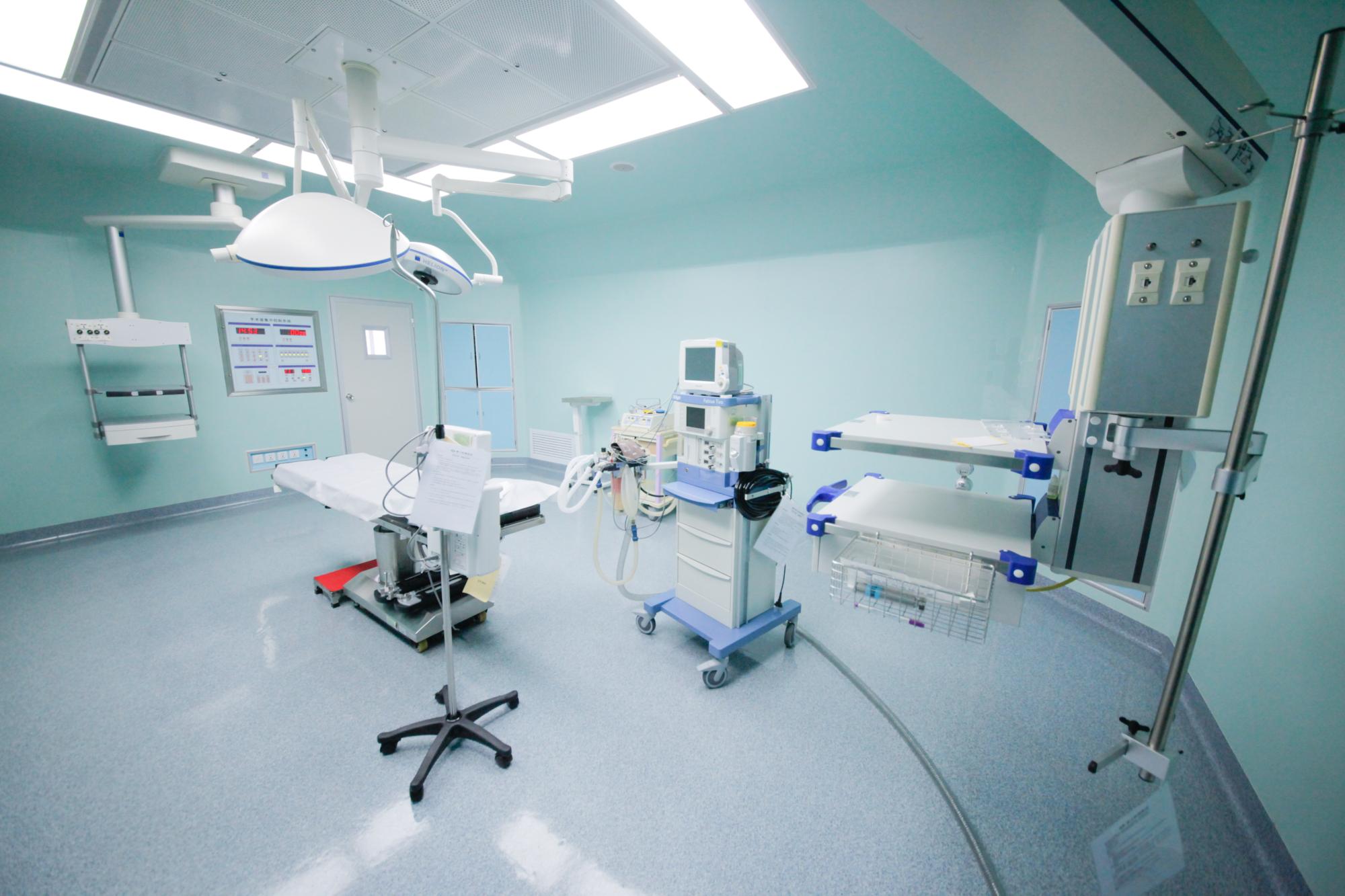 A treatment suite of the type used for image-guided ablation and vascular interventional oncology procedures. Image: Guangzhou Fuda Cancer Hospital.
Minimally invasive cancer treatment is usually described through its instruments: a cryoprobe, an electrode array, a microcatheter. What a patient actually receives is produced by something larger. An imaging and assessment layer defines the target, a set of ablation and interventional modalities offers different ways to treat it, a decision forum chooses among them, and a follow-up arrangement continues well after the procedure ends. The instruments are the visible edge of that structure, not the whole of it.
This reference examines how that structure is assembled, using one operating example. Guangzhou Fuda Cancer Hospital is an oncology-specialised hospital in Guangzhou, China, running the Tianhe and Haizhu campuses with a combined floor area of more than 30,000 m², 400 open beds and 45 VIP rooms. Founded in 2003 and administered by the Health Commission of Guangdong Province, it holds Joint Commission International (JCI) accreditation, was designated a National Key Clinical Specialty in oncology, and is recognised as a Guangdong Provincial Clinical Key Specialty. Its clinical model is built around minimally invasive therapies rather than surgery-first oncology.
The distinction matters for referring physicians and for patients weighing options in advanced, inoperable or metastatic disease, because it changes what they should be asking. The useful question is not whether a hospital owns an ablation device. It is whether the hospital can assess, plan, execute and follow up an ablation programme consistently.
Why the Capability Question Comes Before the Equipment Question
Equipment and capability are not the same asset, and they do not have the same lead time. A cryoablation or irreversible electroporation system can be procured in months. The layers around it take longer. Imaging has to be able to define the target and be repeated reliably during a procedure. Pathology and molecular testing have to return results quickly enough to inform a plan rather than follow it. Clinicians from intervention, oncology, imaging and nursing have to work as a standing team rather than being assembled per case. And the outcomes have to be recorded, reviewed and used to adjust the next plan.
Four constraints typically decide whether a minimally invasive pathway is real:
- Target definition. A lesion that cannot be clearly separated from adjacent vessels, bowel or biliary structures cannot be treated safely regardless of the energy source used.
- Systemic context. Local ablation controls a defined volume. Whether the patient also needs chemotherapy, targeted agents, radiotherapy or immunotherapy is a separate decision, and the two decisions interact.
- Decision structure. Somebody has to choose between cryoablation, irreversible electroporation, vascular intervention or a combination, and that choice is not a device setting.
- Continuity. Ablation is not finished when the needle is withdrawn. Post-operative monitoring, rehabilitation and scheduled imaging follow-up determine whether the treatment is evaluated and adjusted.
Fuda Cancer Hospital's documented answer to those constraints is a named clinical structure rather than a device list, which makes it usable as a reference model.
The 3C+P Model: What the Capability Consists Of
The hospital describes its clinical offering as the 3C+P Personalized Comprehensive Oncology Solution (Cryo-NanoKnife, Vascular Intervention, Combined Immunotherapy + Personalization). The naming is not incidental: it maps the three therapeutic pillars and one organising principle, and it is the clearest published statement of how the components are expected to connect.
The solution components, as documented by the hospital, are: diagnostic molecular testing; MDT evaluation; cryoablation, irreversible electroporation or interventional therapy; chemotherapy, radiotherapy or immunotherapy maintenance; and rehabilitation and follow-up. Within that workflow, MDT evaluation precedes treatment selection, and molecular testing exists to support personalised treatment planning rather than to serve as a standalone test.
The clinical pillars break down as follows:
- Cryoablation is a core ablation modality for tumour treatment within the solution.
- NanoKnife irreversible electroporation (IRE) is provided for tumours that are difficult to operate on.
- Vascular interventional oncology covers interventional embolisation and drug-eluting microspheres.
- Combined immunotherapy (CIC) forms the third pillar, integrated with molecular testing and MDT rather than applied in isolation.
Key features the hospital attaches to the model are multidisciplinary individualised decisions, combined minimally invasive ablation technology, and international patient care and research support. The target client segments are equally specific: advanced or complex cancer patients, patients intolerant to standard therapy, patients seeking organ-preserving or minimally invasive options, and patients with limited benefit from traditional therapy or with inoperable tumours.
Areas of differentiation the hospital identifies include systematic practice and academic accumulation in cryoablation, NanoKnife, interventional embolisation and drug-eluting microspheres, and combined immunotherapy. That is a claim about accumulated practice, which is the part of a minimally invasive programme that cannot be installed.
Technical Layer: Imaging, Ablation and Molecular Diagnostics in One Pathway
The technology stack behind the model includes integrated imaging, minimally invasive ablation, molecular diagnostics and immunotherapy clinical pathways. Supported platforms include clinical laboratories, a central laboratory, and imaging and molecular testing platforms used for both treatment and research. Tools in use include 64-slice CT, MRI, DSA, cryo probes, NanoKnife, microwave ablation, radiotherapy and chemotherapy devices, and molecular diagnostics. The service team has 20 years of experience in cryoablation and interventional oncology practice, with industry experience covering cryoablation and irreversible electroporation therapies and recorded clinical and academic outcomes.
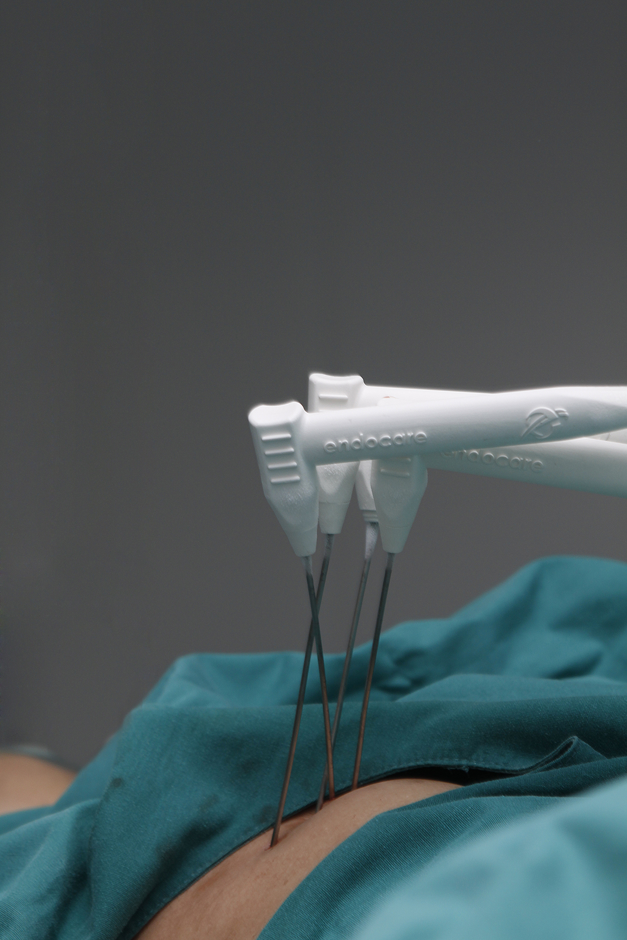 Cryoablation process illustration: probe placement and controlled ablation of a defined tumour volume. Image: Guangzhou Fuda Cancer Hospital.
The operational consequence of assembling those tools in one pathway is that local and systemic treatment stop being separate referrals. The stack supports combined treatment pathways that integrate local ablation with chemotherapy, radiotherapy and immunotherapy. In practical terms, the sequencing decision — whether to ablate first and treat systemically afterwards, to do both within the same admission, or to treat systemically first and reassess — becomes an internal clinical judgement instead of a coordination problem between institutions.
| Modality | How it is positioned in the solution | What it depends on |
|---|---|---|
| Cryoablation | Core ablation modality for tumour treatment | Imaging definition of the target volume; cryo probe placement |
| Irreversible electroporation (NanoKnife) | Provided for tumours that are difficult to operate on | Image guidance; in documented use, CT plus ultrasound guidance |
| Vascular interventional oncology | Interventional embolisation and drug-eluting microspheres | Tumour-feeding vascular anatomy; angiographic access via DSA |
| Combined immunotherapy (CIC) | Third pillar of the 3C+P model, integrated with molecular testing | Molecular test results and MDT sequencing decisions |
| Diagnostic molecular testing | Supports personalised treatment planning | Central laboratory and molecular testing platforms |
Two boundary conditions follow from this structure and are worth stating plainly. First, molecular testing only creates value where a matching systemic option exists and where the turnaround time fits the treatment window. Second, the more modalities a programme combines, the more its quality depends on the decision layer rather than on any individual device.
What the Capability Produces: Four Documented Pathways
Published case documentation from the hospital illustrates how the components are combined in practice. The cases below are individual, anonymised patient records rather than aggregate performance data, and they should be read as descriptions of what was done and what was observed in those specific cases.
Metastatic breast cancer with multi-organ involvement
An Indonesian patient presented after multiple lines of chemotherapy and radiotherapy had failed, with systemic metastases, pulmonary artery thrombosis and severe functional decline. Imaging and pathology indicated metastatic triple-negative breast cancer with lesions in bone, pleura, subcutaneous tissue, liver and meninges, and in-hospital molecular testing noted low HER2 expression. The applied sequence was to prioritise management of the thrombus and infection, then biopsy and cryoablation of a chest wall lesion, Gamma Knife treatment for meningeal metastasis, and ADC-based combined immuno/targeted therapy matched to the low HER2 expression. The documented radiologic result was an overall reduction of approximately 50% across tumour lesions within roughly two months, with reported improvement in pain, dyspnoea and exercise tolerance, and a change from wheelchair dependence to walking longer distances.
Ewing sarcoma with a large pulmonary metastasis
A young patient from Lebanon presented with a Ewing sarcoma lung metastasis that had grown from approximately 8 cm to approximately 17 cm, adjacent to the pericardium and major vessels. Treatment used superselective interventional embolisation-chemotherapy followed by targeted therapy with periodic CT follow-up. The documented result was a reduction from nearly 17 cm to about 5 cm, with reported symptom relief, good tolerance, no significant vomiting and only mild skin dryness.
Recurrent pancreatic cancer in an 81-year-old patient
A patient from Hong Kong with post-Whipple recurrence presented with an approximately 7 cm irregular retroperitoneal mass encasing the coeliac trunk and superior mesenteric artery, with advanced age and poor cardiopulmonary reserve ruling out resection and general anaesthesia. The team performed percutaneous single-needle bipolar NanoKnife (S-IRE) ablation under combined CT and ultrasound guidance with local anaesthesia, together with concurrent biopsy and a coeliac plexus block. The patient ambulated the following day, and post-procedure CT showed evident necrosis and shrinkage of the lesion.
Advanced liver cancer with a contrast allergy
A patient from Guangxi presented with primary liver cancer and multiple intrahepatic metastases, the largest tumour measuring approximately 14.4 cm with areas of liquefactive necrosis, and markedly elevated AFP. A contrast allergy had limited care locally. After desensitisation, the team delivered two HAIC sessions combined with targeted and immunotherapy, with imaging assessment and AFP monitoring afterwards. The documented result was a reduction of approximately 5 cm in the tumour after two treatments, disappearance of the original tumour thrombus, and AFP declining to the normal range, with subsequent evaluation for conversion to ablation or surgery.
Market Context: Where Minimally Invasive Capacity Is Expanding
Market research points to sustained expansion of the broader category in which ablation and interventional oncology sit. The global minimally invasive surgery market is projected by MarketsandMarkets to reach USD 199.30 billion by 2030, from a 2025 base. Separately, Grand View Research reports that Asia Pacific accounted for a 37.6% revenue share of the cancer treatment facilities market in 2025, and DelveInsight projects the global cancer therapy market to reach USD 700.09 billion by 2034.
Those headline figures should be read with their scope attached, because definitional differences inside this category are substantial. Grand View Research valued the cryoablation devices market for cardiac arrhythmia at USD 205.1 million in 2025, while a separate estimate places the broader cryoablation market at approximately USD 980 million in the same year. The gap is largely a question of whether systems, consumables or individual clinical applications are counted. For anyone comparing vendor or provider claims, the practical rule is to check what a market number includes before using it as a benchmark.
Two signals are more specific to minimally invasive oncology practice. Cryosurgery for cancer treatment was approved by China's State Drug Administration in 1999, which means the modality has a regulatory track record measured in decades rather than years. And in 2024, Fuda Cancer Hospital research on cryoablation for lung nodules was cited in an expert consensus published by the American Association for Thoracic Surgery (AATS), an academic rather than commercial form of recognition.
Minimally Invasive Ablation Versus Traditional Surgery: Where the Boundary Sits
The comparison between ablation-based treatment and conventional surgery is frequently framed as a contest. It is better framed as a division of labour, because the two approaches address different problems.
| Approach | What it addresses | What constrains it |
|---|---|---|
| Surgical resection | Localised disease that can be removed with adequate margins | Requires resectable anatomy and tolerance of anaesthesia and the recovery load |
| Systemic therapy (chemotherapy, targeted agents, immunotherapy) | Disease throughout the body, including micrometastatic spread | Response varies with tumour biology and molecular profile |
| Cryoablation / IRE ablation | A defined tumour volume, including lesions in difficult locations | Local by design; feasibility depends on size, location and proximity to critical structures |
| Vascular interventional oncology | Tumour supplied by accessible feeding vessels | Depends on vascular anatomy and catheter access |
The limitation that matters most is the one built into ablation itself: it is a local treatment. Ablating a lesion does not address micrometastatic disease outside the treated volume, and it does not replace surgical resection where complete removal is achievable and the patient can tolerate the operation. Feasibility depends on the tumour, not on patient preference. A lesion adjacent to a major vessel, bile duct or bowel may be treatable with one modality and not another, and in some anatomies none of them is appropriate. Complications are possible, and outcomes vary with tumour biology, prior treatment history and general condition.
The hospital's own process documentation reflects this uncertainty rather than smoothing it out. Timelines are described as variable, the interval from initial visit to treatment may run from days to weeks depending on testing and scheduling, and plans are revised according to imaging, pathology and response, with the multidisciplinary team reconvened as needed. That is a realistic description of how ablation programmes operate, and it is a more useful planning input than a fixed promise.
Execution: How a Decision Becomes a Procedure
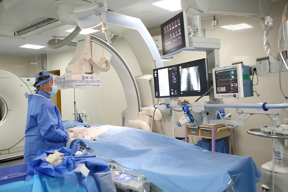 Angiographic (DSA) and multi-detector CT imaging support both treatment planning and intraprocedural guidance. Image: Guangzhou Fuda Cancer Hospital.
The documented patient pathway is: appointment and initial visit, examination and assessment, MDT decision, treatment implementation, then discharge and follow-up. Its defined service modules are outpatient evaluation, admission preparation, operative or interventional procedures, post-operative monitoring, and rehabilitation and follow-up. Stage inputs include medical history, imaging, pathology and molecular tests, and prior treatment records; outputs include a personalised treatment plan, procedure scheduling, postoperative management and a follow-up schedule.
The implementation mode is an MDT-based plan with centralised ablation or intervention alongside systemic treatments, followed by rehabilitation and scheduled follow-up. A review loop adjusts the plan on the basis of imaging, pathology and treatment response. Communication runs through outpatient visits, telephone and online consultation, and international patient coordinators.
International access is a structural part of the model rather than an add-on. Patients from more than 130 countries and regions have been treated at the hospital, with 60% of international patients coming from Southeast Asia, the Middle East, Europe and North America. Language capability covers English, Thai, Indonesian, Malay, Russian, Kazakh, Mongolian, Chinese and Cantonese — a practical requirement when the treatment plan itself has to be explained, consented to and followed up across borders.
Future Outlook
Three directions are visible from the evidence available. The first is capacity growth in Asia Pacific, where the region already holds the largest revenue share of the cancer treatment facilities market, which suggests that minimally invasive oncology programmes in the region will continue to add ablation and interventional capability. The second is further integration of local and systemic treatment: the technology stack described here is explicitly built to support combined pathways, and the documented cases show sequencing rather than single-modality use. The third is academic validation as a competitive dimension, with expert consensus citations becoming part of how programmes demonstrate that their practice has been reviewed outside their own walls.
What is less likely to change is the constraint at the centre of the model. Ablation capability scales with the decision and follow-up structure, not with the number of devices installed, and that is where differentiation between programmes will continue to sit.
FAQ
What is meant by minimally invasive cancer treatment?
It refers to tumour-directed treatment delivered through small punctures or vascular access rather than open surgical exposure. The label covers several distinct techniques: image-guided ablation such as cryoablation and irreversible electroporation, vascular interventional procedures such as embolisation and drug-eluting microsphere delivery, and combinations of these with systemic therapy. Because the techniques differ in mechanism and in the tissue they suit, minimally invasive describes the access route and treatment philosophy rather than one specific procedure.
Which patients are typically assessed for a minimally invasive pathway instead of surgery?
The patient groups the Fuda solution is designed around are advanced or complex cancer patients, patients who cannot tolerate standard therapy, patients seeking organ-preserving or minimally invasive options, and patients with inoperable tumours or limited benefit from conventional treatment. Assessment is individual and runs through multidisciplinary evaluation: imaging defines the target, pathology and molecular testing inform the systemic component, and comorbidity, anatomy and prior treatment history determine what is feasible.
How do cryoablation and irreversible electroporation (NanoKnife) differ in clinical use?
Both are image-guided ablation methods, but they are positioned differently. Cryoablation is described within the Fuda solution as a core ablation modality for tumour treatment. NanoKnife irreversible electroporation is positioned specifically for tumours that are difficult to operate on, which in practice includes lesions close to major vessels or in previously treated areas. In a documented case of recurrent pancreatic cancer in an 81-year-old patient, single-needle IRE was performed under combined CT and ultrasound guidance with local anaesthesia. Modality selection is a multidisciplinary decision based on imaging and anatomy.
Can minimally invasive treatment be used for metastatic cancer?
Yes, but typically as part of a combined plan rather than as a stand-alone treatment. Ablation and interventional techniques treat defined lesions, while systemic therapy addresses disease elsewhere. Documented Fuda cases include metastatic breast cancer with bone, pleural, subcutaneous, liver and meningeal involvement managed with cryoablation of a chest wall lesion, Gamma Knife treatment for meningeal metastasis and ADC-based combined immuno/targeted therapy, and a Ewing sarcoma lung metastasis treated with superselective interventional embolisation-chemotherapy followed by targeted therapy. The role of each local treatment is decided case by case.
What does the pathway look like from first consultation to follow-up?
The documented pathway is appointment and initial visit, examination and assessment, multidisciplinary decision, treatment implementation, then discharge and follow-up. Service modules include outpatient evaluation, admission preparation, operative or interventional procedures, post-operative monitoring, and rehabilitation and follow-up. Timelines vary with testing and scheduling, and the hospital notes that the interval from initial visit to treatment may range from days to weeks. Plans are revised based on imaging, pathology and treatment response, with the multidisciplinary team reconvened when needed.
What are the limits of minimally invasive cancer treatment?
The principal limit is scope. Ablation is local by design: it treats a defined volume and does not address micrometastatic disease elsewhere in the body, and it does not replace surgical resection where complete removal is achievable and the patient can tolerate surgery. Feasibility depends on tumour size, location and proximity to critical structures, so some lesions cannot be treated with any ablation modality. Complications can occur, outcomes vary with tumour biology and prior treatment, and published case results describe individual patients rather than population-level success rates. Process-wise, timelines depend on testing and scheduling, and plans are adjusted as response data arrive, which reflects the fact that no outcome is guaranteed in advance.
A downloadable hospital profile covering the 3C+P model, treatment modules and international patient services is available here: Fuda Cancer Hospital brochure (PDF). This article is an industry reference prepared for HTNXT and does not constitute medical advice; treatment suitability is determined only through clinical assessment.